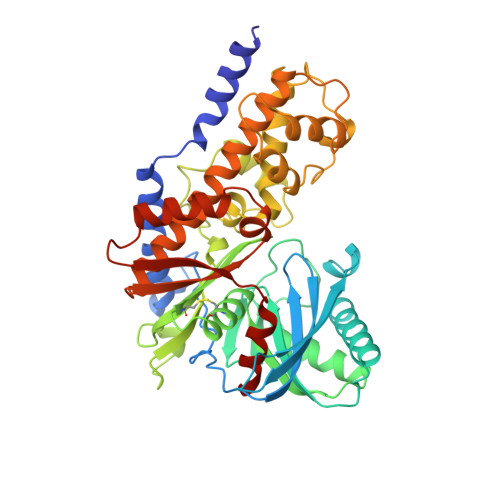
1BDG

HEXOKINASE FROM SCHISTOSOMA MANSONI COMPLEXED WITH GLUCOSE


Experimental Data Snapshot

  • Method: X-RAY DIFFRACTION
  • Resolution: 2.60 Å
  • R-Value Free: 0.256 
  • R-Value Work: 0.173 
  • R-Value Observed: 0.173 

Starting Model: experimental
View more details

wwPDB Validation   3D Report Full Report


Ligand Structure Quality Assessment 


This is version 1.5 of the entry. See complete history


Literature

The structure of mammalian hexokinase-1.

Mulichak, A.M.Wilson, J.E.Padmanabhan, K.Garavito, R.M.

(1998) Nat Struct Biol 5: 555-560

  • DOI: https://doi.org/10.1038/811
  • Primary Citation of Related Structures:  
    1BDG, 1BG3

  • PubMed Abstract: 

    We have determined the structures of the glucose-6-phosphate (G6P)-inhibitable 100,000 Mr Type I hexokinase from rat and the G6P-sensitive 50,000 Mr hexokinase from Schistosoma mansoni at a resolution of 2.8 and 2.6 A respectively. The structures define the glucose and G6P binding sites in these enzymes, suggest the mechanisms of intradomain G6P inhibition and activity loss in the Type I hexokinase N-terminal half, and reveal the structure of the membrane targeting motif that integrates the Type I hexokinase into the outer mitochondrial membrane.


  • Organizational Affiliation

    Department of Biochemistry, Michigan State University, East Lansing, USA. mulichak@alecto.bch.msu.edu


Macromolecules
Find similar proteins by:  (by identity cutoff)  |  3D Structure
Entity ID: 1
MoleculeChains Sequence LengthOrganismDetailsImage
HEXOKINASE451Schistosoma mansoniMutation(s): 0 
EC: 2.7.1.1
UniProt
Find proteins for Q26609 (Schistosoma mansoni)
Explore Q26609 
Go to UniProtKB:  Q26609
Entity Groups  
Sequence Clusters30% Identity50% Identity70% Identity90% Identity95% Identity100% Identity
UniProt GroupQ26609
Sequence Annotations
Expand
  • Reference Sequence
Experimental Data & Validation

Experimental Data

  • Method: X-RAY DIFFRACTION
  • Resolution: 2.60 Å
  • R-Value Free: 0.256 
  • R-Value Work: 0.173 
  • R-Value Observed: 0.173 
  • Space Group: P 61 2 2
Unit Cell:
Length ( Å )Angle ( ˚ )
a = 114.3α = 90
b = 114.3β = 90
c = 155.2γ = 120
Software Package:
Software NamePurpose
R-AXISdata collection
R-AXISdata reduction
AMoREphasing
X-PLORrefinement
R-AXISdata scaling

Structure Validation

View Full Validation Report


Ligand Structure Quality Assessment 


Entry History 

Deposition Data

Revision History  (Full details and data files)

  • Version 1.0: 1999-05-11
    Type: Initial release
  • Version 1.1: 2008-03-24
    Changes: Version format compliance
  • Version 1.2: 2011-07-13
    Changes: Version format compliance
  • Version 1.3: 2020-07-29
    Type: Remediation
    Reason: Carbohydrate remediation
    Changes: Data collection, Derived calculations, Structure summary
  • Version 1.4: 2022-12-21
    Changes: Database references, Structure summary
  • Version 1.5: 2023-08-09
    Changes: Refinement description