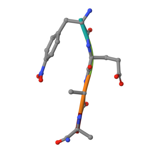
1YTJ

SIV PROTEASE CRYSTALLIZED WITH PEPTIDE PRODUCT


Experimental Data Snapshot

  • Method: X-RAY DIFFRACTION
  • Resolution: 2.50 Å
  • R-Value Free: 0.285 
  • R-Value Work: 0.174 
  • R-Value Observed: 0.174 

wwPDB Validation   3D Report Full Report


This is version 1.3 of the entry. See complete history


Literature

Three-dimensional structures of HIV-1 and SIV protease product complexes.

Rose, R.B.Craik, C.S.Douglas, N.L.Stroud, R.M.

(1996) Biochemistry 35: 12933-12944

  • DOI: https://doi.org/10.1021/bi9612733
  • Primary Citation of Related Structures:  
    1YTG, 1YTH, 1YTI, 1YTJ

  • PubMed Abstract: 

    Strain is eliminated as a factor in hydrolysis of the scissile peptide bond by human immunodeficiency virus (HIV)-1 and simian immunodeficiency virus (SIV), based on the first eight complexes of products of hydrolysis with the enzymes. The carboxyl group generated at the scissile bond interacts with both catalytic aspartic acids. The structures directly suggest the interactions of the gemdiol intermediate with the active site. Based on the structures, the nucleophilic water is displaced stereospecifically by substrate binding toward one catalytic aspartic acid, while the scissile carbonyl becomes hydrogen bonded to the other catalytic aspartic acid in position for hydrolysis. Crystal structures for two N-terminal (P) products and two C-terminal (Q) products provide unambiguous density for the ligands at 2.2-2.6 A resolution and 17-21% R factors. The N-terminal product, Ac-S-L-N-F/, overlaps closely with the N-terminal sequences of peptidomimetic inhibitors bound to the protease. Comparison of the two C-terminal products, /F-L-E-K and /F(NO2)-E-A-Nle-S, indicates that the P2' residue is highly constrained, while the positioning of the P1' and P3' residues are sequence dependent.


  • Organizational Affiliation

    Department of Biochemistry and Biophysics, University of California, San Francisco 94143-0448, USA.


Macromolecules
Find similar proteins by:  (by identity cutoff)  |  3D Structure
Entity ID: 1
MoleculeChains Sequence LengthOrganismDetailsImage
SIV PROTEASE99Simian immunodeficiency virusMutation(s): 1 
Gene Names: SIV(MAC)239
EC: 3.4.23.16
UniProt
Find proteins for P05896 (Simian immunodeficiency virus (isolate Mm142-83))
Explore P05896 
Go to UniProtKB:  P05896
Entity Groups  
Sequence Clusters30% Identity50% Identity70% Identity90% Identity95% Identity100% Identity
UniProt GroupP05896
Sequence Annotations
Expand
  • Reference Sequence

Find similar proteins by:  Sequence   |   3D Structure  

Entity ID: 2
MoleculeChains Sequence LengthOrganismDetailsImage
PEPTIDE PRODUCTB [auth I]5N/AMutation(s): 0 
Sequence Annotations
Expand
  • Reference Sequence
Small Molecules
Modified Residues  2 Unique
IDChains TypeFormula2D DiagramParent
NLE
Query on NLE
B [auth I]L-PEPTIDE LINKINGC6 H13 N O2LEU
PPN
Query on PPN
B [auth I]L-PEPTIDE LINKINGC9 H10 N2 O4PHE
Experimental Data & Validation

Experimental Data

  • Method: X-RAY DIFFRACTION
  • Resolution: 2.50 Å
  • R-Value Free: 0.285 
  • R-Value Work: 0.174 
  • R-Value Observed: 0.174 
  • Space Group: C 2 2 21
Unit Cell:
Length ( Å )Angle ( ˚ )
a = 62.3α = 90
b = 32.1β = 90
c = 96.3γ = 90
Software Package:
Software NamePurpose
X-PLORmodel building
X-PLORrefinement
DENZOdata reduction
X-PLORphasing

Structure Validation

View Full Validation Report


Entry History 

Deposition Data

Revision History  (Full details and data files)

  • Version 1.0: 1997-03-12
    Type: Initial release
  • Version 1.1: 2008-03-24
    Changes: Version format compliance
  • Version 1.2: 2011-07-13
    Changes: Atomic model, Database references, Derived calculations, Non-polymer description, Structure summary, Version format compliance
  • Version 1.3: 2021-11-03
    Changes: Advisory, Database references, Derived calculations, Other