Structural mechanism for ubiquitinated-cargo recognition by the Golgi-localized, {gamma}-ear-containing, ADP-ribosylation-factor-binding proteins
Prag, G., Lee, S., Mattera, R., Arighi, C.N., Beach, B.M., Bonifacino, J.S., Hurley, J.H.(2005) Proc Natl Acad Sci U S A 102: 2334-2339
- PubMed: 15701688 Search on PubMedSearch on PubMed Central
- DOI: https://doi.org/10.1073/pnas.0500118102
- Primary Citation Related Structures:
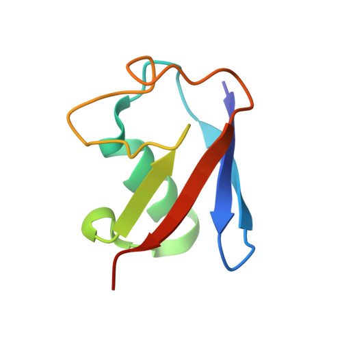
1YD8 - PubMed Abstract:
The Golgi-localized, gamma-ear-containing, Arf (ADP-ribosylation factor)-binding (GGA) proteins are clathrin adaptors that mediate the sorting of transmembrane-cargo molecules at the trans-Golgi network and endosomes. Cargo proteins can be directed into the GGA pathway by at least two different types of sorting signals: acidic cluster-dileucine motifs and covalent modification by ubiquitin. The latter modification is recognized by the GGAs through binding to their GAT [GGA and TOM (target of Myb)] domain. Here we report the crystal structure of the GAT domain of human GGA3 in a 1:1 complex with ubiquitin at 2.8-A resolution. Ubiquitin binds to a hydrophobic and acidic patch on helices alpha1 and alpha2 of the GAT three-helix bundle that includes Asn-223, Leu-227, Glu-230, Met-231, Asp-244, Glu-246, Leu-247, Glu-250, and Leu-251. The GAT-binding surface on ubiquitin is a hydrophobic patch centered on Ile-44 that is also responsible for binding most other ubiquitin effectors. The ubiquitin-binding site observed in the crystal is distinct from the Rabaptin-5-binding site on helices alpha2 and alpha3 of the GAT domain. Mutational analysis and modeling of the ubiquitin-Rabaptin-5-GAT ternary complex indicates that ubiquitin and Rabaptin-5 can bind to the GAT domain at two different sites without any steric conflict. This ability highlights the GAT domain as a hub for interactions with multiple partners in trafficking.
- Laboratory of Molecular Biology, National Institute of Diabetes and Digestive and Kidney Diseases, National Institutes of Health, Bethesda, MD 20892, USA.
Organizational Affiliation: