1Y42 | pdb_00001y42

Crystal structure of a C-terminally truncated CYT-18 protein


Experimental Data Snapshot

  • Method: X-RAY DIFFRACTION
  • Resolution: 1.95 Å
  • R-Value Free: 
    0.235 (Depositor), 0.236 (DCC) 
  • R-Value Work: 
    0.174 (Depositor), 0.182 (DCC) 
  • R-Value Observed: 
    0.179 (Depositor) 

Starting Model: experimental
View more details

wwPDB Validation 3D Report Full Report

Validation slider image for 1Y42

Ligand Structure Quality Assessment 


This is version 1.4 of the entry. See complete history

Literature

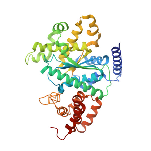
A tyrosyl-tRNA synthetase adapted to function in group I intron splicing by acquiring a new RNA binding surface.

Paukstelis, P.J.Coon, R.Madabusi, L.Nowakowski, J.Monzingo, A.Robertus, J.Lambowitz, A.M.

(2005) Mol Cell 17: 417-428

  • DOI: https://doi.org/10.1016/j.molcel.2004.12.026
  • Primary Citation Related Structures: 
    1Y42

  • PubMed Abstract: 

    We determined a 1.95 A X-ray crystal structure of a C-terminally truncated Neurospora crassa mitochondrial tyrosyl-tRNA synthetase (CYT-18 protein) that functions in splicing group I introns. CYT-18's nucleotide binding fold and intermediate alpha-helical domains superimpose on those of bacterial TyrRSs, except for an N-terminal extension and two small insertions not found in nonsplicing bacterial enzymes. These additions surround the cyt-18-1 mutation site and are sites of suppressor mutations that restore splicing, but not synthetase activity. Highly constrained models based on directed hydroxyl radical cleavage assays show that the group I intron binds at a site formed in part by the three additions on the nucleotide binding fold surface opposite that which binds tRNATyr. Our results show how essential proteins can progressively evolve new functions.


  • Organizational Affiliation
    • Institute for Cellular and Molecular Biology, Department of Chemistry and Biochemistry and Section of Molecular Genetics and Microbiology, School of Biological Sciences, University of Texas, Austin, TX 78712, USA.

Macromolecule Content 

  • Total Structure Weight: 45.1 kDa 
  • Atom Count: 3,161 
  • Modeled Residue Count: 370 
  • Deposited Residue Count: 392 
  • Unique protein chains: 1

Macromolecules

Find similar proteins by:|  3D Structure
Entity ID: 1
MoleculeChains  Sequence LengthOrganismDetailsImage
Tyrosyl-tRNA synthetase, mitochondrialA [auth X]392Neurospora crassaMutation(s): 0 
Gene Names: cyt-18
EC: 6.1.1.1
UniProt
Find proteins for P12063 (Neurospora crassa (strain ATCC 24698 / 74-OR23-1A / CBS 708.71 / DSM 1257 / FGSC 987))
Explore P12063 
Go to UniProtKB:  P12063
Entity Groups
Sequence Clusters30% Identity50% Identity70% Identity90% Identity95% Identity100% Identity
UniProt GroupP12063
Sequence Annotations
Expand
Reference Sequence

Small Molecules

Ligands 1 Unique
IDChains Name / Formula / InChI Key2D Diagram3D Interactions
TYR

Query on TYR


Download:Ideal Coordinates CCD File
B [auth X]TYROSINE
C9 H11 N O3
OUYCCCASQSFEME-QMMMGPOBSA-N

Experimental Data & Validation

Experimental Data

  • Method: X-RAY DIFFRACTION
  • Resolution: 1.95 Å
  • R-Value Free:  0.235 (Depositor), 0.236 (DCC) 
  • R-Value Work:  0.174 (Depositor), 0.182 (DCC) 
  • R-Value Observed: 0.179 (Depositor) 
Space Group: C 1 2 1
Unit Cell:
Length ( Å )Angle ( ˚ )
a = 104.88α = 90
b = 73.21β = 111.35
c = 56.79γ = 90
Software Package:
Software NamePurpose
REFMACrefinement
MAR345data collection
SCALEPACKdata scaling
CNSphasing

Structure Validation

View Full Validation Report


Ligand Structure Quality Assessment 


Entry History 

Deposition Data

Revision History  (Full details and data files)

  • Version 1.0: 2005-02-15
    Type: Initial release
  • Version 1.1: 2008-04-30
    Changes: Version format compliance
  • Version 1.2: 2011-07-13
    Changes: Derived calculations, Version format compliance
  • Version 1.3: 2017-10-11
    Changes: Refinement description
  • Version 1.4: 2023-08-23
    Changes: Data collection, Database references, Derived calculations, Refinement description