Plasticity, hydration and accessibility in ribonuclease A. The structure of a new crystal form and its low-humidity variant.
Sadasivan, C., Nagendra, H.G., Vijayan, M.(1998) Acta Crystallogr D Biol Crystallogr 54: 1343-1352
- PubMed: 10089510 Search on PubMed
- DOI: https://doi.org/10.1107/s0907444998005149
- Primary Citation Related Structures:
1XPS, 1XPT - PubMed Abstract:
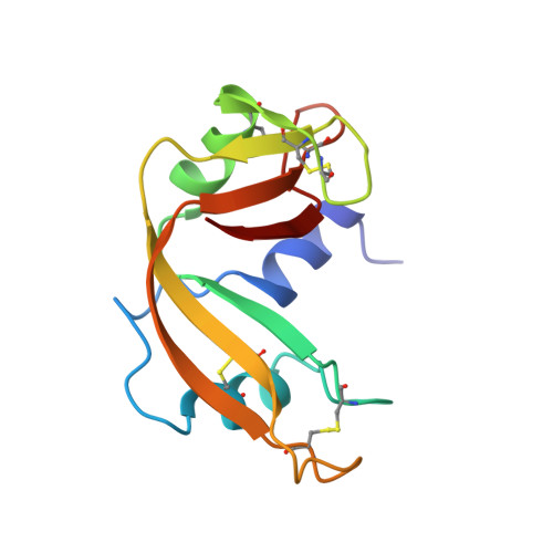
The structures of a new crystal form of ribonuclease A and its low-humidity variant, each containing two crystallographically independent molecules, have been determined and refined. A detailed comparison of these structures with those of the other known crystal forms of the enzyme, which have different packing arrangements and solvent composition, leads to a meaningful delineation of the rigid and flexible regions of the protein molecule and the nature of its plasticity. Many of the water molecules which are common to all the structures are involved in bridging different regions of the protein molecule, thus emphasizing the role of water in stabilizing the tertiary structure. The analysis of the structures shows that for a given N or O atom, the level of hydration increases with accessible surface area, but levels off at an area of about 10 A2. Generally, the hydration level tends to drop when the area increases beyond about 20 A2. This drop correlates with an increase in the displacement parameter. The analysis also suggests that the van der Waals radii and probe radius normally used in accessible surface area calculations are not appropriate for dealing with all situations.
- Molecular Biophysics Unit, Indian Institute of Science, Bangalore 560 012, India.
Organizational Affiliation: