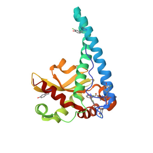
1XIL

HYDROGEN BONDING IN HUMAN MANGANESE SUPEROXIDE DISMUTASE CONTAINING 3-FLUOROTYROSINE


Experimental Data Snapshot

  • Method: X-RAY DIFFRACTION
  • Resolution: 1.53 Å

Starting Model: experimental
View more details

wwPDB Validation   3D Report Full Report


This is version 1.5 of the entry. See complete history


Literature

Hydrogen bonding in human manganese superoxide dismutase containing 3-fluorotyrosine

Ayala, I.Perry, J.J.Szczepanski, J.Tainer, J.A.Vala, M.T.Nick, H.S.Silverman, D.N.

(2005) Biophys J 89: 4171-4179

  • DOI: https://doi.org/10.1529/biophysj.105.060616
  • Primary Citation of Related Structures:  
    1XDC, 1XIL

  • PubMed Abstract: 

    Incorporation of 3-fluorotyrosine and site-specific mutagenesis has been utilized with Fourier transform infrared (FTIR) spectroscopy and x-ray crystallography to elucidate active-site structure and the role of an active-site residue Tyr34 in human manganese superoxide dismutase (MnSOD). Calculated harmonic frequencies at the B3LYP/6-31G** level of theory for L-tyrosine and its 3-fluorine substituted analog are compared to experimental frequencies for vibrational mode assignments. Each of the nine tyrosine residues in each of the four subunits of the homotetramer of human MnSOD was replaced with 3-fluorotyrosine. The crystal structures of the unfluorinated and fluorinated wild-type MnSOD are nearly superimposable with the root mean-square deviation for 198 alpha-carbon atoms at 0.3 A. The FTIR data show distinct vibrational modes arising from 3-fluorotyrosine in MnSOD. Comparison of spectra for wild-type and Y34F MnSOD showed that the phenolic hydroxyl of Tyr34 is hydrogen bonded, acting as a proton donor in the active site. Comparison with crystal structures demonstrates that the hydroxyl of Tyr34 is a hydrogen bond donor to an adjacent water molecule; this confirms the participation of Tyr34 in a network of residues and water molecules that extends from the active site to the adjacent subunit.


  • Organizational Affiliation

    Department of Pharmacology, University of Florida, Gainesville, Florida 32610, USA.


Macromolecules
Find similar proteins by:  (by identity cutoff)  |  3D Structure
Entity ID: 1
MoleculeChains Sequence LengthOrganismDetailsImage
Superoxide dismutase [Mn], mitochondrial
A, B
198Homo sapiensMutation(s): 9 
Gene Names: SOD2
EC: 1.15.1.1
UniProt & NIH Common Fund Data Resources
Find proteins for P04179 (Homo sapiens)
Explore P04179 
Go to UniProtKB:  P04179
PHAROS:  P04179
Entity Groups  
Sequence Clusters30% Identity50% Identity70% Identity90% Identity95% Identity100% Identity
UniProt GroupP04179
Sequence Annotations
Expand
  • Reference Sequence
Small Molecules
Modified Residues  1 Unique
IDChains TypeFormula2D DiagramParent
YOF
Query on YOF
A, B
L-PEPTIDE LINKINGC9 H10 F N O3TYR
Experimental Data & Validation

Experimental Data

  • Method: X-RAY DIFFRACTION
  • Resolution: 1.53 Å
  • Space Group: P 21 21 2
Unit Cell:
Length ( Å )Angle ( ˚ )
a = 73.696α = 90
b = 75.309β = 90
c = 67.992γ = 90
Software Package:
Software NamePurpose
SHELXL-97refinement
SCALEPACKdata scaling
CNSrefinement
HKL-2000data collection
HKL-2000data reduction
AMoREphasing

Structure Validation

View Full Validation Report


Entry History 

Deposition Data

Revision History  (Full details and data files)

  • Version 1.0: 2005-03-08
    Type: Initial release
  • Version 1.1: 2008-04-30
    Changes: Version format compliance
  • Version 1.2: 2011-07-13
    Changes: Derived calculations, Version format compliance
  • Version 1.3: 2019-07-24
    Changes: Data collection, Derived calculations, Refinement description
  • Version 1.4: 2021-10-20
    Changes: Database references, Derived calculations
  • Version 1.5: 2023-08-23
    Changes: Data collection, Refinement description