Role of water in plasticity, stability, and action of proteins: the crystal structures of lysozyme at very low levels of hydration.
Nagendra, H.G., Sukumar, N., Vijayan, M.(1998) Proteins 32: 229-240
- PubMed: 9714162 Search on PubMed
- DOI: https://doi.org/10.1002/(sici)1097-0134(19980801)32:2<229::aid-prot9>3.0.co;2-f
- Primary Citation Related Structures:
1XEI, 1XEJ, 1XEK - PubMed Abstract:
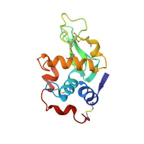
Earlier studies involving water-mediated transformations in lysozyme and ribonuclease A have shown that the overall movements in the protein molecule consequent to the reduction in the amount of surrounding water are similar to those that occur during enzyme action, thus highlighting the relationship among hydration, plasticity, and action of these enzymes. Monoclinic lysozyme retains its crystallinity even when the level of hydration is reduced further below that necessary for activity (about 0.2 gram of water per gram of protein). In order to gain insights into the role of water in the stability and the plasticity of the protein molecule and the geometrical basis for the loss of activity that accompanies dehydration, the crystal structures of monoclinic lysozyme with solvent contents of 17.6%, 16.9%, and 9.4% were determined and refined. A detailed comparison of these forms with the normally hydrated forms show that the C-terminal segment (residues 88-129) of domain I and the main loop (residues 65-73) in domain II exhibit large deviations in atomic positions when the solvent content is reduced, although the three-dimensional structure is essentially preserved. Many crucial water bridges between different regions of the molecule are conserved in spite of differences in detail, even when the level of hydration is reduced well below that required for activity. The loss of activity that accompany dehydration appears to be caused by the removal of functionally important water molecules from the active-site region and the reduction in the size of the substrate binding cleft.
- Molecular Biophysics Unit, Indian Institute of Science, Bangalore.
Organizational Affiliation: