1WNX | pdb_00001wnx

D136E mutant of Heme Oxygenase from Corynebacterium diphtheriae (HmuO)


Experimental Data Snapshot

  • Method: X-RAY DIFFRACTION
  • Resolution: 1.85 Å
  • R-Value Free: 
    0.201 (Depositor), 0.208 (DCC) 
  • R-Value Work: 
    0.150 (Depositor), 0.165 (DCC) 
  • R-Value Observed: 
    0.155 (Depositor) 

wwPDB Validation 3D Report Full Report

Validation slider image for 1WNX

Ligand Structure Quality Assessment 


This is version 1.4 of the entry. See complete history

Literature

Roles of Distal Asp in Heme Oxygenase from Corynebacterium diphtheriae, HmuO: A WATER-DRIVEN OXYGEN ACTIVATION MECHANISM

Matsui, T.Furukawa, M.Unno, M.Tomita, T.Ikeda-Saito, M.

(2005) J Biological Chem 280: 2981-2989

  • DOI: https://doi.org/10.1074/jbc.M410263200
  • Primary Citation Related Structures: 
    1WNV, 1WNW, 1WNX

  • PubMed Abstract: 

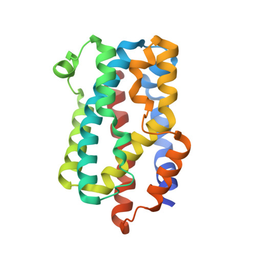
    Heme oxygenases found in mammals, plants, and bacteria catalyze degradation of heme using the same mechanism. Roles of distal Asp (Asp-136) residue in HmuO, a heme oxygenase of Corynebacterium diphtheriae, have been investigated by site-directed mutagenesis, enzyme kinetics, resonance Raman spectroscopy, and x-ray crystallography. Replacements of the Asp-136 by Ala and Phe resulted in reduced heme degradation activity due to the formation of ferryl heme, showing that the distal Asp is critical in HmuO heme oxygenase activity. D136N HmuO catalyzed heme degradation at a similar efficiency to wild type and D136E HmuO, implying that the carboxylate moiety is not required for the heme catabolism by HmuO. Resonance Raman results suggest that the inactive ferryl heme formation in the HmuO mutants is induced by disruption of the interaction between a reactive Fe-OOH species and an adjacent distal pocket water molecule. Crystal structural analysis of the HmuO mutants confirms partial disappearance of this nearby water in D136A HmuO. Our results provide the first experimental evidence for the catalytic importance of the nearby water molecule that can be universally critical in heme oxygenase catalysis and propose that the distal Asp helps in positioning the key water molecule at a position suitable for efficient activation of the Fe-OOH species.


  • Organizational Affiliation
    • Institute of Multidisciplinary Research for Advanced Materials, Tohoku University, Katahira, Aoba, Sendai 980-8577, Japan.

Macromolecule Content 

  • Total Structure Weight: 50.68 kDa 
  • Atom Count: 3,835 
  • Modeled Residue Count: 417 
  • Deposited Residue Count: 430 
  • Unique protein chains: 1

Macromolecules

Find similar proteins by:|  3D Structure
Entity ID: 1
MoleculeChains  Sequence LengthOrganismDetailsImage
Heme oxygenase
A, B
215Corynebacterium diphtheriaeMutation(s): 1 
EC: 1.14.99.3 (PDB Primary Data), 1.14.14.18 (UniProt)
UniProt
Find proteins for P71119 (Corynebacterium diphtheriae (strain ATCC 700971 / NCTC 13129 / Biotype gravis))
Explore P71119 
Go to UniProtKB:  P71119
Entity Groups
Sequence Clusters30% Identity50% Identity70% Identity90% Identity95% Identity100% Identity
UniProt GroupP71119
Sequence Annotations
Expand
Reference Sequence

Small Molecules

Ligands 3 Unique
IDChains Name / Formula / InChI Key2D Diagram3D Interactions
HEM

Query on HEM


Download:Ideal Coordinates CCD File
I [auth A],
P [auth B]
PROTOPORPHYRIN IX CONTAINING FE
C34 H32 Fe N4 O4
KABFMIBPWCXCRK-RGGAHWMASA-L
SO4

Query on SO4


Download:Ideal Coordinates CCD File
C [auth A]
D [auth A]
E [auth A]
F [auth A]
G [auth A]
C [auth A],
D [auth A],
E [auth A],
F [auth A],
G [auth A],
J [auth B],
K [auth B],
L [auth B],
M [auth B],
N [auth B],
O [auth B]
SULFATE ION
O4 S
QAOWNCQODCNURD-UHFFFAOYSA-L
NA

Query on NA


Download:Ideal Coordinates CCD File
H [auth A]SODIUM ION
Na
FKNQFGJONOIPTF-UHFFFAOYSA-N

Experimental Data & Validation

Experimental Data

  • Method: X-RAY DIFFRACTION
  • Resolution: 1.85 Å
  • R-Value Free:  0.201 (Depositor), 0.208 (DCC) 
  • R-Value Work:  0.150 (Depositor), 0.165 (DCC) 
  • R-Value Observed: 0.155 (Depositor) 
Space Group: C 1 2 1
Unit Cell:
Length ( Å )Angle ( ˚ )
a = 106.151α = 90
b = 63.722β = 130.28
c = 79.296γ = 90
Software Package:
Software NamePurpose
REFMACrefinement
HKL-2000data reduction
SCALEPACKdata scaling
CNSphasing

Structure Validation

View Full Validation Report


Ligand Structure Quality Assessment 


Entry History 

Deposition Data

Revision History  (Full details and data files)

  • Version 1.0: 2004-11-09
    Type: Initial release
  • Version 1.1: 2008-04-30
    Changes: Version format compliance
  • Version 1.2: 2011-07-13
    Changes: Version format compliance
  • Version 1.3: 2021-11-10
    Changes: Database references, Derived calculations
  • Version 1.4: 2024-05-29
    Changes: Data collection