1WE1 | pdb_00001we1

Crystal structure of heme oxygenase-1 from cyanobacterium Synechocystis sp. PCC6803 in complex with heme


Experimental Data Snapshot

  • Method: X-RAY DIFFRACTION
  • Resolution: 2.50 Å
  • R-Value Free: 
    0.269 (Depositor), 0.256 (DCC) 
  • R-Value Work: 
    0.220 (Depositor), 0.211 (DCC) 
  • R-Value Observed: 
    0.224 (Depositor) 

Starting Model: experimental
View more details

wwPDB Validation 3D Report Full Report

Validation slider image for 1WE1

Ligand Structure Quality Assessment 


This is version 1.3 of the entry. See complete history

Literature

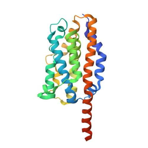
Crystal structure of heme oxygenase-1 from cyanobacterium Synechocystis sp. PCC 6803 in complex with heme

Sugishima, M.Migita, C.T.Zhang, X.Yoshida, T.Fukuyama, K.

(2004) Eur J Biochem 271: 4517-4525

  • DOI: https://doi.org/10.1111/j.1432-1033.2004.04411.x
  • Primary Citation Related Structures: 
    1WE1

  • PubMed Abstract: 

    Heme oxygenase (HO) catalyzes the oxidative degradation of heme utilizing molecular oxygen and reducing equivalents. In photosynthetic organisms, HO functions in the biosynthesis of such open-chain tetrapyrroles as phyto-chromobilin and phycobilins, which are involved in the signal transduction for light responses and light harvesting for photosynthesis, respectively. We have determined the first crystal structure of a HO-1 from a photosynthetic organism, Synechocystis sp. PCC 6803 (Syn HO-1), in complex with heme at 2.5 A resolution. Heme-Syn HO-1 shares a common folding with other heme-HOs. Although the heme pocket of heme-Syn HO-1 is, for the most part, similar to that of mammalian HO-1, they differ in such features as the flexibility of the distal helix and hydrophobicity. In addition, 2-propanol derived from the crystallization solution occupied the hydrophobic cavity, which is proposed to be a CO trapping site in rat HO-1 that suppresses product inhibition. Although Syn HO-1 and mammalian HO-1 are similar in overall structure and amino acid sequence (57% similarity vs. human HO-1), their molecular surfaces differ in charge distribution. The surfaces of the heme binding sides are both positively charged, but this patch of Syn HO-1 is narrow compared to that of mammalian HO-1. This feature is suited to the selective binding of ferredoxin, the physiological redox partner of Syn HO-1; the molecular size of ferredoxin is approximately 10 kDa whereas the size of NADPH-cytochrome P450 reductase, a reducing partner of mammalian HO-1, is approximately 77 kDa. A docking model of heme-Syn HO-1 and ferredoxin suggests indirect electron transfer from an iron-sulfur cluster in ferredoxin to the heme iron of heme-Syn HO-1.


  • Organizational Affiliation
    • Department of Biology, Graduate School of Science, Osaka University, Toyonaka, Osaka, Japan.

Macromolecule Content 

  • Total Structure Weight: 111.65 kDa 
  • Atom Count: 7,468 
  • Modeled Residue Count: 887 
  • Deposited Residue Count: 960 
  • Unique protein chains: 1

Macromolecules

Find similar proteins by:|  3D Structure
Entity ID: 1
MoleculeChains  Sequence LengthOrganismDetailsImage
Heme oxygenase 1
A, B, C, D
240Synechocystis sp. PCC 6803Mutation(s): 0 
EC: 1.14.99.3 (PDB Primary Data), 1.14.14.18 (UniProt)
UniProt
Find proteins for P72849 (Synechocystis sp. (strain ATCC 27184 / PCC 6803 / Kazusa))
Explore P72849 
Go to UniProtKB:  P72849
Entity Groups
Sequence Clusters30% Identity50% Identity70% Identity90% Identity95% Identity100% Identity
UniProt GroupP72849
Sequence Annotations
Expand
Reference Sequence

Small Molecules

Ligands 4 Unique
IDChains Name / Formula / InChI Key2D Diagram3D Interactions
HEM

Query on HEM


Download:Ideal Coordinates CCD File
H [auth A],
M [auth B],
Q [auth C],
U [auth D]
PROTOPORPHYRIN IX CONTAINING FE
C34 H32 Fe N4 O4
KABFMIBPWCXCRK-RGGAHWMASA-L
PO4

Query on PO4


Download:Ideal Coordinates CCD File
E [auth A],
K [auth B]
PHOSPHATE ION
O4 P
NBIIXXVUZAFLBC-UHFFFAOYSA-K
IPA

Query on IPA


Download:Ideal Coordinates CCD File
I [auth A]
J [auth A]
N [auth B]
O [auth B]
R [auth C]
I [auth A],
J [auth A],
N [auth B],
O [auth B],
R [auth C],
S [auth C],
V [auth D],
W [auth D]
ISOPROPYL ALCOHOL
C3 H8 O
KFZMGEQAYNKOFK-UHFFFAOYSA-N
CL

Query on CL


Download:Ideal Coordinates CCD File
F [auth A],
G [auth A],
L [auth B],
P [auth C],
T [auth D]
CHLORIDE ION
Cl
VEXZGXHMUGYJMC-UHFFFAOYSA-M

Experimental Data & Validation

Experimental Data

  • Method: X-RAY DIFFRACTION
  • Resolution: 2.50 Å
  • R-Value Free:  0.269 (Depositor), 0.256 (DCC) 
  • R-Value Work:  0.220 (Depositor), 0.211 (DCC) 
  • R-Value Observed: 0.224 (Depositor) 
Space Group: C 1 2 1
Unit Cell:
Length ( Å )Angle ( ˚ )
a = 110.79α = 90
b = 113.73β = 112.26
c = 109.7γ = 90
Software Package:
Software NamePurpose
MOSFLMdata reduction
SCALAdata scaling
MOLREPphasing
CNSrefinement
CCP4data scaling

Structure Validation

View Full Validation Report


Ligand Structure Quality Assessment 


Entry History 

Deposition Data

Revision History  (Full details and data files)

  • Version 1.0: 2004-12-21
    Type: Initial release
  • Version 1.1: 2008-04-30
    Changes: Version format compliance
  • Version 1.2: 2011-07-13
    Changes: Source and taxonomy, Version format compliance
  • Version 1.3: 2023-10-25
    Changes: Data collection, Database references, Derived calculations, Refinement description