1W78

E.coli FolC in complex with DHPP and ADP


Experimental Data Snapshot

  • Method: X-RAY DIFFRACTION
  • Resolution: 1.82 Å
  • R-Value Free: 0.203 
  • R-Value Work: 0.165 

wwPDB Validation   3D Report Full Report


This is version 1.3 of the entry. See complete history


Literature

Escherichia Coli Folc Structure Reveals an Unexpected Dihydrofolate Binding Site Providing an Attractive Target for Anti-Microbial Therapy

Mathieu, M.Debousker, G.Vincent, S.Viviani, F.Bamas-Jacques, N.Mikol, V.

(2005) J Biol Chem 280: 18916

  • DOI: https://doi.org/10.1074/jbc.M413799200
  • Primary Citation of Related Structures:  
    1W78, 1W7K

  • PubMed Abstract: 

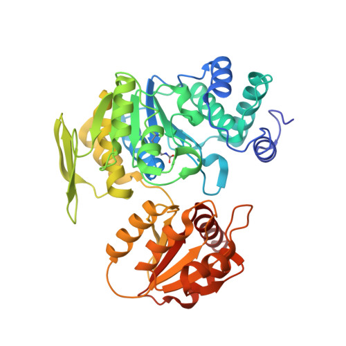
    In some bacteria, such as Escherichia coli, the addition of L-glutamate to dihydropteroate (dihydrofolate synthetase activity) and the subsequent additions of L-glutamate to tetrahydrofolate (folylpolyglutamate synthetase (FPGS) activity) are catalyzed by the same enzyme, FolC. The crystal structure of E. coli FolC is described in this paper. It showed strong similarities to that of the FPGS enzyme of Lactobacillus casei within the ATP binding site and the catalytic site, as do all other members of the Mur synthethase superfamily. FolC structure revealed an unexpected dihydropteroate binding site very different from the folate site identified previously in the FPGS structure. The relevance of this site is exemplified by the presence of phosphorylated dihydropteroate, a reaction intermediate in the DHFS reaction. L. casei FPGS is considered a relevant model for human FPGS. As such, the presence of a folate binding site in E. coli FolC, which is different from the one seen in FPGS enzymes, provides avenues for the design of specific inhibitors of this enzyme in antimicrobial therapy.


  • Organizational Affiliation

    Department of Structural Biology, Aventis Pharma, 13 Quai J. Guesde, F-94403 Vitry/Seine, France. magali.mathieu@sanofi-aventis.com


Macromolecules
Find similar proteins by:  (by identity cutoff)  |  3D Structure
Entity ID: 1
MoleculeChains Sequence LengthOrganismDetailsImage
FOLC BIFUNCTIONAL PROTEIN422Escherichia coliMutation(s): 0 
EC: 6.3.2.12
UniProt
Find proteins for P08192 (Escherichia coli (strain K12))
Explore P08192 
Go to UniProtKB:  P08192
Entity Groups  
Sequence Clusters30% Identity50% Identity70% Identity90% Identity95% Identity100% Identity
UniProt GroupP08192
Sequence Annotations
Expand
  • Reference Sequence
Small Molecules
Ligands 4 Unique
IDChains Name / Formula / InChI Key2D Diagram3D Interactions
ADP
Query on ADP

Download Ideal Coordinates CCD File 
F [auth A]ADENOSINE-5'-DIPHOSPHATE
C10 H15 N5 O10 P2
XTWYTFMLZFPYCI-KQYNXXCUSA-N
PD8
Query on PD8

Download Ideal Coordinates CCD File 
B [auth A]PHOSPHORYLATED DIHYDROPTEROATE
C14 H13 N6 O6 P
DNKVHXNVVYQUDC-UHFFFAOYSA-N
SO4
Query on SO4

Download Ideal Coordinates CCD File 
E [auth A]SULFATE ION
O4 S
QAOWNCQODCNURD-UHFFFAOYSA-L
MG
Query on MG

Download Ideal Coordinates CCD File 
C [auth A],
D [auth A]
MAGNESIUM ION
Mg
JLVVSXFLKOJNIY-UHFFFAOYSA-N
Modified Residues  1 Unique
IDChains TypeFormula2D DiagramParent
KCX
Query on KCX
A
L-PEPTIDE LINKINGC7 H14 N2 O4LYS
Experimental Data & Validation

Experimental Data

  • Method: X-RAY DIFFRACTION
  • Resolution: 1.82 Å
  • R-Value Free: 0.203 
  • R-Value Work: 0.165 
  • Space Group: P 21 21 21
Unit Cell:
Length ( Å )Angle ( ˚ )
a = 64.368α = 90
b = 80.62β = 90
c = 92.829γ = 90
Software Package:
Software NamePurpose
BUSTERrefinement
MOSFLMdata reduction
SCALAdata scaling
AMoREphasing

Structure Validation

View Full Validation Report


Entry History 

Deposition Data

Revision History  (Full details and data files)

  • Version 1.0: 2005-02-09
    Type: Initial release
  • Version 1.1: 2011-05-08
    Changes: Version format compliance
  • Version 1.2: 2011-07-13
    Changes: Version format compliance
  • Version 1.3: 2023-12-13
    Changes: Data collection, Database references, Derived calculations, Refinement description