1W0Y | pdb_00001w0y

tf7a_3771 complex


Experimental Data Snapshot

  • Method: X-RAY DIFFRACTION
  • Resolution: 2.50 Å
  • R-Value Free: 
    0.258 (Depositor), 0.202 (DCC) 
  • R-Value Work: 
    0.199 (Depositor) 

Starting Model: experimental
View more details

wwPDB Validation 3D Report Full Report

Validation slider image for 1W0Y

Ligand Structure Quality Assessment 


This is version 1.4 of the entry. See complete history

Literature

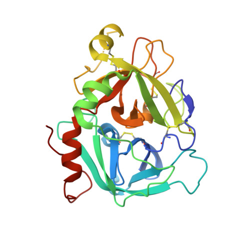
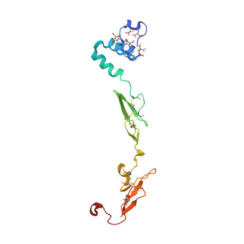
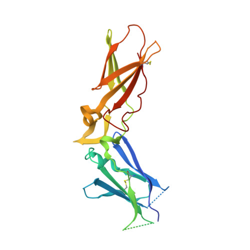
Design of Selective Phenylglycine Amide Tissue Factor/Factor Viia Inhibitors

Groebke-Zbinden, K.Banner, D.W.Ackermann, J.D'Arcy, A.Kirchhofer, D.Ji, Y.-H.Tschopp, T.B.Wallbaum, S.Weber, L.

(2005) Bioorg Med Chem Lett 15: 817

  • DOI: https://doi.org/10.1016/j.bmcl.2004.10.092
  • Primary Citation Related Structures: 
    1W0Y, 1W2K

  • PubMed Abstract: 

    Proof of concept experiments have shown that tissue factor/factor VIIa inhibitors have antithrombotic activity without enhancing bleeding propensity. Starting from lead compounds generated by a biased combinatorial approach, phenylglycine amide tissue factor/factor VIIa inhibitors with low nanomolar affinity and good selectivity against other serine proteases of the coagulation cascade were designed, using the guidance of X-ray structural analysis and molecular modelling.


  • Organizational Affiliation
    • F. Hoffmann-La Roche Ltd, CH-4070 Basel, Switzerland. katrin.groebke_zbinden@roche.com

Macromolecule Content 

  • Total Structure Weight: 69.48 kDa 
  • Atom Count: 5,060 
  • Modeled Residue Count: 581 
  • Deposited Residue Count: 606 
  • Unique protein chains: 3

Macromolecules

Find similar proteins by:|  3D Structure
Entity ID: 1
MoleculeChains  Sequence LengthOrganismDetailsImage
BLOOD COAGULATION FACTOR VIIAA [auth H]254Homo sapiensMutation(s): 0 
EC: 3.4.21.21
UniProt & NIH Common Fund Data Resources
Find proteins for P08709 (Homo sapiens)
Explore P08709 
Go to UniProtKB:  P08709
PHAROS:  P08709
GTEx:  ENSG00000057593 
Entity Groups
Sequence Clusters30% Identity50% Identity70% Identity90% Identity95% Identity100% Identity
UniProt GroupP08709
Sequence Annotations
Expand
Reference Sequence
Find similar proteins by:|  3D Structure
Entity ID: 2
MoleculeChains  Sequence LengthOrganismDetailsImage
BLOOD COAGULATION FACTOR VIIAB [auth L]142Homo sapiensMutation(s): 0 
EC: 3.4.21.21
UniProt & NIH Common Fund Data Resources
Find proteins for P08709 (Homo sapiens)
Explore P08709 
Go to UniProtKB:  P08709
PHAROS:  P08709
GTEx:  ENSG00000057593 
Entity Groups
Sequence Clusters30% Identity50% Identity70% Identity90% Identity95% Identity100% Identity
UniProt GroupP08709
Glycosylation
Glycosylation Sites: 2Go to GlyGen: P08709-1
Sequence Annotations
Expand
Reference Sequence
Find similar proteins by:|  3D Structure
Entity ID: 3
MoleculeChains  Sequence LengthOrganismDetailsImage
TISSUE FACTORC [auth T]210Homo sapiensMutation(s): 0 
UniProt & NIH Common Fund Data Resources
Find proteins for P13726 (Homo sapiens)
Explore P13726 
Go to UniProtKB:  P13726
PHAROS:  P13726
GTEx:  ENSG00000117525 
Entity Groups
Sequence Clusters30% Identity50% Identity70% Identity90% Identity95% Identity100% Identity
UniProt GroupP13726
Sequence Annotations
Expand
Reference Sequence

Small Molecules

Ligands 5 Unique
IDChains Name / Formula / InChI Key2D Diagram3D Interactions
771

Query on 771


Download:Ideal Coordinates CCD File
D [auth H]4-(4-BENZYLOXY-2-METHANESULFONYLAMINO-5-METHOXY-BENZYLAMINO)-BENZAMIDINE
C23 H26 N4 O4 S
CRFICMUDNBIMKL-UHFFFAOYSA-N
BGC

Query on BGC


Download:Ideal Coordinates CCD File
P [auth L]beta-D-glucopyranose
C6 H12 O6
WQZGKKKJIJFFOK-VFUOTHLCSA-N
FUC

Query on FUC


Download:Ideal Coordinates CCD File
G [auth L]alpha-L-fucopyranose
C6 H12 O5
SHZGCJCMOBCMKK-SXUWKVJYSA-N
CAC

Query on CAC


Download:Ideal Coordinates CCD File
F [auth H]CACODYLATE ION
C2 H6 As O2
OGGXGZAMXPVRFZ-UHFFFAOYSA-M
CA

Query on CA


Download:Ideal Coordinates CCD File
E [auth H]
H [auth L]
I [auth L]
J [auth L]
K [auth L]
E [auth H],
H [auth L],
I [auth L],
J [auth L],
K [auth L],
L,
M [auth L],
N [auth L],
O [auth L]
CALCIUM ION
Ca
BHPQYMZQTOCNFJ-UHFFFAOYSA-N
Modified Residues  1 Unique
IDChains TypeFormula2D DiagramParent
CGU
Query on CGU
B [auth L]L-PEPTIDE LINKINGC6 H9 N O6GLU
Binding Affinity Annotations 
IDSourceBinding Affinity
771 BindingDB:  1W0Y Ki: 380 (nM) from 1 assay(s)

Experimental Data & Validation

Experimental Data

  • Method: X-RAY DIFFRACTION
  • Resolution: 2.50 Å
  • R-Value Free:  0.258 (Depositor), 0.202 (DCC) 
  • R-Value Work:  0.199 (Depositor) 
Space Group: P 21 21 21
Unit Cell:
Length ( Å )Angle ( ˚ )
a = 71.338α = 90
b = 82.378β = 90
c = 123.963γ = 90
Software Package:
Software NamePurpose
CNXrefinement
XDSdata reduction
XDSdata scaling
AMoREphasing

Structure Validation

View Full Validation Report


Ligand Structure Quality Assessment 


Entry History 

Deposition Data

Revision History  (Full details and data files)

  • Version 1.0: 2005-01-21
    Type: Initial release
  • Version 1.1: 2011-05-08
    Changes: Version format compliance
  • Version 1.2: 2011-07-13
    Changes: Version format compliance
  • Version 1.3: 2020-07-29
    Type: Remediation
    Reason: Carbohydrate remediation
    Changes: Advisory, Data collection, Derived calculations, Other, Structure summary
  • Version 1.4: 2023-12-13
    Changes: Advisory, Data collection, Database references, Refinement description, Structure summary