1W0V

Crystal Structure Of HLA-B*2705 Complexed With the self-Peptide TIS from EGF-response factor 1


Experimental Data Snapshot

  • Method: X-RAY DIFFRACTION
  • Resolution: 2.27 Å
  • R-Value Free: 0.246 
  • R-Value Work: 0.184 
  • R-Value Observed: 0.187 

wwPDB Validation   3D Report Full Report


This is version 1.2 of the entry. See complete history


Literature

Thermodynamic and Structural Equivalence of Two Hla-B27 Subtypes Complexed with a Self-Peptide

Hulsmeyer, M.Welfle, K.Pohlmann, T.Misselwitz, R.Alexiev, U.Welfle, H.Saenger, W.Uchanska-Ziegler, B.Ziegler, A.

(2005) J Mol Biol 346: 1367

  • DOI: https://doi.org/10.1016/j.jmb.2004.12.047
  • Primary Citation of Related Structures:  
    1W0V, 1W0W

  • PubMed Abstract: 

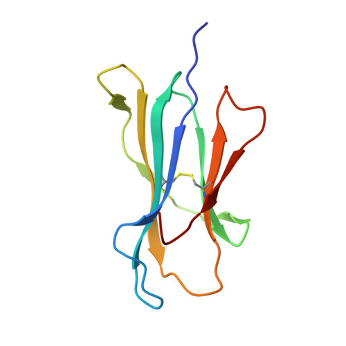
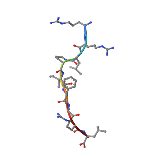
    The F pocket of major histocompatibility complex (in humans HLA) class I molecules accommodates the C terminus of the bound peptide. Residues forming this pocket exhibit considerable polymorphism, and a single difference (Asp116 in HLA-B*2705 and His116 in HLA-B*2709 heavy chains) confers differential association of these two HLA-B27 subtypes to the autoimmune disease ankylosing spondylitis. As peptide presentation by HLA molecules is of central importance for immune responses, we performed thermodynamic (circular dichroism, differential scanning calorimetry, fluorescence polarization) and X-ray crystallographic analyses of both HLA-B27 subtypes complexed with the epidermal growth factor response factor 1-derived self-peptide TIS (RRLPIFSRL) to understand the impact of the Asp116His exchange on peptide display. This peptide is known to be presented in vivo by both subtypes, and as expected for a self-peptide, TIS-reactive cytotoxic T lymphocytes are absent in the respective individuals. The thermodynamic analyses reveal that both HLA-B27:TIS complexes exhibit comparable, relatively high thermostability (Tm approximately 60 degrees C) and undergo multi-step unfolding reactions, with dissociation of the peptide in the first step. As shown by X-ray crystallography, only subtle structural differences between the subtypes were observed regarding the architecture of their F pockets, including the presence of distinct networks of water molecules. However, no consistent structural differences were found between the peptide presentation modes. In contrast to other peptides displayed by the two HLA-subtypes which show either structural or dynamical differences in their peptide presentation modes, the TIS-complexed HLA-B*2705 and HLA-B*2709 subtypes are an example for thermodynamic and structural equivalence, in agreement with functional data.


  • Organizational Affiliation

    Institut für Chemie/Kristallographie, Freie Universität Berlin, Takustrasse 6, 14195 Berlin, Germany.


Macromolecules
Find similar proteins by:  (by identity cutoff)  |  3D Structure
Entity ID: 1
MoleculeChains Sequence LengthOrganismDetailsImage
HLA CLASS I HISTOCOMPATIBILITY ANTIGEN276Homo sapiensMutation(s): 0 
UniProt & NIH Common Fund Data Resources
Find proteins for P01889 (Homo sapiens)
Explore P01889 
Go to UniProtKB:  P01889
PHAROS:  P01889
GTEx:  ENSG00000234745 
Entity Groups  
Sequence Clusters30% Identity50% Identity70% Identity90% Identity95% Identity100% Identity
UniProt GroupP01889
Sequence Annotations
Expand
  • Reference Sequence
Find similar proteins by:  (by identity cutoff)  |  3D Structure
Entity ID: 2
MoleculeChains Sequence LengthOrganismDetailsImage
BETA-2-MICROGLOBULIN100Homo sapiensMutation(s): 0 
UniProt & NIH Common Fund Data Resources
Find proteins for P61769 (Homo sapiens)
Explore P61769 
Go to UniProtKB:  P61769
PHAROS:  P61769
GTEx:  ENSG00000166710 
Entity Groups  
Sequence Clusters30% Identity50% Identity70% Identity90% Identity95% Identity100% Identity
UniProt GroupP61769
Sequence Annotations
Expand
  • Reference Sequence

Find similar proteins by:  Sequence   |   3D Structure  

Entity ID: 3
MoleculeChains Sequence LengthOrganismDetailsImage
BUTYRATE RESPONSE FACTOR 29Homo sapiensMutation(s): 0 
UniProt & NIH Common Fund Data Resources
Find proteins for P47974 (Homo sapiens)
Explore P47974 
Go to UniProtKB:  P47974
PHAROS:  P47974
GTEx:  ENSG00000152518 
Entity Groups  
UniProt GroupP47974
Sequence Annotations
Expand
  • Reference Sequence
Experimental Data & Validation

Experimental Data

  • Method: X-RAY DIFFRACTION
  • Resolution: 2.27 Å
  • R-Value Free: 0.246 
  • R-Value Work: 0.184 
  • R-Value Observed: 0.187 
  • Space Group: P 21 21 21
Unit Cell:
Length ( Å )Angle ( ˚ )
a = 50.861α = 90
b = 82.488β = 90
c = 109.421γ = 90
Software Package:
Software NamePurpose
REFMACrefinement
HKLdata reduction
SCALEPACKdata scaling
MOLREPphasing

Structure Validation

View Full Validation Report


Entry History 

Deposition Data

Revision History  (Full details and data files)

  • Version 1.0: 2005-03-07
    Type: Initial release
  • Version 1.1: 2011-07-13
    Changes: Advisory, Version format compliance
  • Version 1.2: 2023-12-13
    Changes: Data collection, Database references, Other, Refinement description