Atomic Resolution Structures and Solution Behavior of Enzyme-Substrate Complexes of Enterobacter Cloacae Pb2 Pentaerythritol Tetranitrate Reductase: Multiple Conformational States and Implications for the Mechanism of Nitroaromatic Explosive Degradation
Khan, H., Barna, T., Harris, R., Bruce, N., Barsukov, I., Munro, A., Moody, P.C.E., Scrutton, N.(2004) J Biol Chem 279: 30563
- PubMed: 15128738 Search on PubMed
- DOI: https://doi.org/10.1074/jbc.M403541200
- Primary Citation Related Structures:
1VYP, 1VYR, 1VYS - PubMed Abstract:
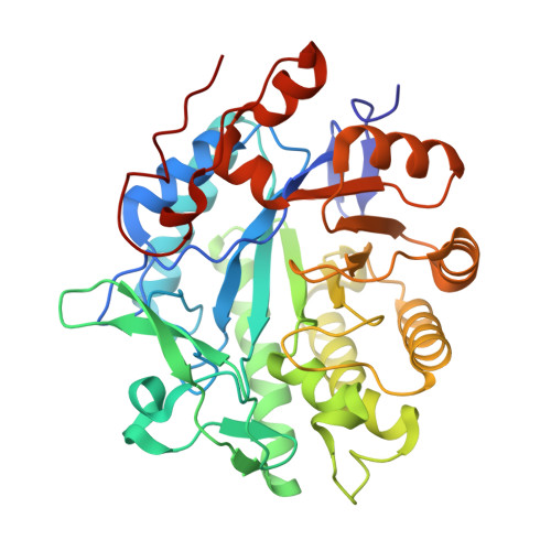
The structure of pentaerythritol tetranitrate (PETN) reductase in complex with the nitroaromatic substrate picric acid determined previously at 1.55 A resolution indicated additional electron density between the indole ring of residue Trp-102 and the nitro group at C-6 of picrate. The data suggested the presence of an unusual bond between substrate and the tryptophan side chain. Herein, we have extended the resolution of the PETN reductase-picric acid complex to 0.9 A. This high-resolution analysis indicates that the active site is partially occupied with picric acid and that the anomalous density seen in the original study is attributed to the population of multiple conformational states of Trp-102 and not a formal covalent bond between the indole ring of Trp-102 and picric acid. The significance of any interaction between Trp-102 and nitroaromatic substrates was probed further in solution and crystal complexes with wild-type and mutant (W102Y and W102F) enzymes. Unlike with wild-type enzyme, in the crystalline form picric acid was bound at full occupancy in the mutant enzymes, and there was no evidence for multiple conformations of active site residues. Solution studies indicate tighter binding of picric acid in the active sites of the W102Y and W102F enzymes. Mutation of Trp-102 does not impair significantly enzyme reduction by NADPH, but the kinetics of decay of the hydride-Meisenheimer complex are accelerated in the mutant enzymes. The data reveal that decay of the hydride-Meisenheimer complex is enzyme catalyzed and that the final distribution of reaction products for the mutant enzymes is substantially different from wild-type enzyme. Implications for the mechanism of high explosive degradation by PETN reductase are discussed.
- Department of Biochemistry, University of Leicester, University Road, Leicester LE1 7RH, United Kingdom.
Organizational Affiliation: