Solution structure and backbone dynamics of component IV Glycera dibranchiata monomeric hemoglobin-CO.
Volkman, B.F., Alam, S.L., Satterlee, J.D., Markley, J.L.(1998) Biochemistry 37: 10906-10919
- PubMed: 9692983 Search on PubMed
- DOI: https://doi.org/10.1021/bi980810b
- Primary Citation Related Structures:
1VRE, 1VRF - PubMed Abstract:
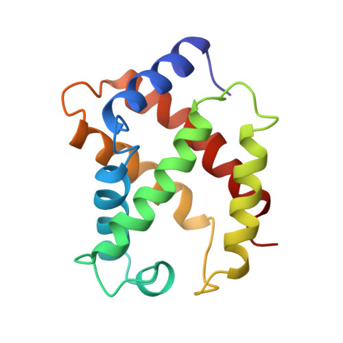
The solution structure and backbone dynamics of the recombinant, ferrous CO-ligated form of component IV monomeric hemoglobin from Glycera dibranchiata (GMH4CO) have been characterized by NMR spectroscopy. Distance geometry and simulated annealing calculations utilizing a total of 2550 distance and torsion angle constraints yielded an ensemble of 29 structures with an overall average backbone rmsd of 0.48 A from the average structure. Differences between the solution structure and a related crystal structure are confined to regions of lower precision in either the NMR or X-ray structure, or in regions where the amino acid sequences differ. 15N relaxation measurements at 76.0 and 60.8 MHz were analyzed with an extended model-free approach, and revealed low-frequency motions in the vicinity of the heme, concentrated in the F helix. Amide proton protection factors were obtained from H-D amide exchange measurements on 15N-labeled protein. Patterns in the backbone dynamics and protection factors were shown to correlate with regions of heterogeneity and disorder in the ensemble of NMR structures and with large crystallographic B-factors in the X-ray structures. Surprisingly, while the backbone atoms of the F helix have higher rmsds and larger measures of dynamics on the microsecond to millisecond time scale than the other helices, amide protection factors for residues in the F helix were observed to be similar to those of the other helices. This contrasts with H-D amide exchange measurements on sperm whale myoglobin which indicated low protection for the F helix (S. N. Loh and B. F. Volkman, unpublished results). These results for GMH4 suggest a model in which the F helix undergoes collective motions as a relatively rigid hydrogen-bonded unit, possibly pivoting about a central position near residue Val87.
- National Magnetic Resonance Facility at Madison, Department of Biochemistry, University of Wisconsin-Madison 53706, USA.
Organizational Affiliation: