


Crystal structure of Ferritin (TM1128) from Thermotoga maritima at 2.00 A resolution
Joint Center for Structural Genomics (JCSG)To be published.
Experimental Data Snapshot
Starting Model: experimental
View more details
wwPDB Validation 3D Report Full Report

Entity ID: 1 | |||||
|---|---|---|---|---|---|
| Molecule | Chains | Sequence Length | Organism | Details | Image |
| ferritin | 176 | Thermotoga maritima MSB8 | Mutation(s): 0 Gene Names: TM1128 EC: 1.16.3.2 | 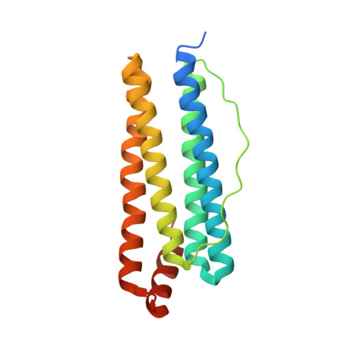 | |
UniProt | |||||
Entity Groups | |||||
| Sequence Clusters | 30% Identity50% Identity70% Identity90% Identity95% Identity100% Identity | ||||
| UniProt Group | Q9X0L2 | ||||
Sequence AnnotationsExpand | |||||
Reference Sequence | |||||
| Ligands 3 Unique | |||||
|---|---|---|---|---|---|
| ID | Chains | Name / Formula / InChI Key | 2D Diagram | 3D Interactions | |
| SO4 Download:Ideal Coordinates CCD File | AA [auth E] BA [auth E] FA [auth F] J [auth A] K [auth A] | SULFATE ION O4 S QAOWNCQODCNURD-UHFFFAOYSA-L |  | ||
| GOL Download:Ideal Coordinates CCD File | CA [auth E] DA [auth E] GA [auth F] HA [auth F] JA [auth G] | GLYCEROL C3 H8 O3 PEDCQBHIVMGVHV-UHFFFAOYSA-N |  | ||
| FE Download:Ideal Coordinates CCD File | EA [auth F] I [auth A] IA [auth G] KA [auth H] N [auth B] | FE (III) ION Fe VTLYFUHAOXGGBS-UHFFFAOYSA-N |  | ||
| Length ( Å ) | Angle ( ˚ ) |
|---|---|
| a = 175.634 | α = 90 |
| b = 175.634 | β = 90 |
| c = 354.843 | γ = 120 |
| Software Name | Purpose |
|---|---|
| REFMAC | refinement |
| SCALA | data scaling |
| CNS | refinement |
| MOLREP | phasing |
| MOSFLM | data reduction |
| CCP4 | data scaling |
| CNS | phasing |


