1VK3 | pdb_00001vk3

Crystal structure of Phosphoribosylformylglycinamidine synthase II (TM1246) from Thermotoga maritima at 2.15 A resolution


Experimental Data Snapshot

  • Method: X-RAY DIFFRACTION
  • Resolution: 2.15 Å
  • R-Value Free: 
    0.254 (Depositor), 0.256 (DCC) 
  • R-Value Work: 
    0.186 (Depositor), 0.190 (DCC) 
  • R-Value Observed: 
    0.190 (Depositor) 

wwPDB Validation 3D Report Full Report

Validation slider image for 1VK3

This is version 1.4 of the entry. See complete history

Literature

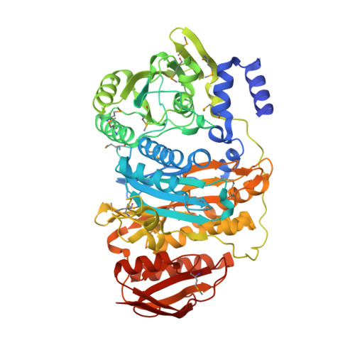
Crystal structure of phosphoribosylformylglycinamidine synthase II (smPurL) from Thermotoga maritima at 2.15 A resolution.

Mathews, I.I.Krishna, S.S.Schwarzenbacher, R.McMullan, D.Abdubek, P.Ambing, E.Canaves, J.M.Chiu, H.J.Deacon, A.M.DiDonato, M.Elsliger, M.A.Godzik, A.Grittini, C.Grzechnik, S.K.Hale, J.Hampton, E.Han, G.W.Haugen, J.Jaroszewski, L.Klock, H.E.Koesema, E.Kreusch, A.Kuhn, P.Lesley, S.A.Levin, I.Miller, M.D.Moy, K.Nigoghossian, E.Paulsen, J.Quijano, K.Reyes, R.Spraggon, G.Stevens, R.C.van den Bedem, H.Velasquez, J.White, A.Wolf, G.Xu, Q.Hodgson, K.O.Wooley, J.Wilson, I.A.

(2006) Proteins 63: 1106-1111

Macromolecule Content 

  • Total Structure Weight: 68.34 kDa 
  • Atom Count: 4,666 
  • Modeled Residue Count: 586 
  • Deposited Residue Count: 615 
  • Unique protein chains: 1

Macromolecules

Find similar proteins by:|  3D Structure
Entity ID: 1
MoleculeChains  Sequence LengthOrganismDetailsImage
Phosphoribosylformylglycinamidine synthase II615Thermotoga maritimaMutation(s): 17 
Gene Names: PURLTM1246
EC: 6.3.5.3
UniProt
Find proteins for Q9X0X3 (Thermotoga maritima (strain ATCC 43589 / DSM 3109 / JCM 10099 / NBRC 100826 / MSB8))
Explore Q9X0X3 
Go to UniProtKB:  Q9X0X3
Entity Groups
Sequence Clusters30% Identity50% Identity70% Identity90% Identity95% Identity100% Identity
UniProt GroupQ9X0X3
Sequence Annotations
Expand
Reference Sequence

Small Molecules

Ligands 1 Unique
IDChains Name / Formula / InChI Key2D Diagram3D Interactions
CL

Query on CL


Download:Ideal Coordinates CCD File
B [auth A]CHLORIDE ION
Cl
VEXZGXHMUGYJMC-UHFFFAOYSA-M
Modified Residues  1 Unique
IDChains TypeFormula2D DiagramParent
MSE
Query on MSE
A
L-PEPTIDE LINKINGC5 H11 N O2 SeMET

Experimental Data & Validation

Experimental Data

  • Method: X-RAY DIFFRACTION
  • Resolution: 2.15 Å
  • R-Value Free:  0.254 (Depositor), 0.256 (DCC) 
  • R-Value Work:  0.186 (Depositor), 0.190 (DCC) 
  • R-Value Observed: 0.190 (Depositor) 
Space Group: P 21 21 21
Unit Cell:
Length ( Å )Angle ( ˚ )
a = 59.775α = 90
b = 72.689β = 90
c = 128.418γ = 90
Software Package:
Software NamePurpose
MOSFLMdata reduction
SCALAdata scaling
SOLVEphasing
RESOLVEmodel building
REFMACrefinement
CCP4data scaling
RESOLVEphasing

Structure Validation

View Full Validation Report


Entry History 

Deposition Data

Revision History  (Full details and data files)

  • Version 1.0: 2004-05-11
    Type: Initial release
  • Version 1.1: 2008-04-26
    Changes: Version format compliance
  • Version 1.2: 2011-07-13
    Changes: Version format compliance
  • Version 1.3: 2023-01-25
    Changes: Database references, Derived calculations
  • Version 1.4: 2024-11-20
    Changes: Data collection, Structure summary