1VFE

HUMAN LACTOFERRIN, N-TERMINAL LOBE MUTANT WITH ARG 121 REPLACED BY SER (R121S)


Experimental Data Snapshot

  • Method: X-RAY DIFFRACTION
  • Resolution: 2.30 Å
  • R-Value Free: 0.270 
  • R-Value Work: 0.196 

wwPDB Validation   3D Report Full Report


This is version 1.5 of the entry. See complete history


Literature

Mutation of arginine 121 in lactoferrin destabilizes iron binding by disruption of anion binding: crystal structures of R121S and R121E mutants.

Faber, H.R.Baker, C.J.Day, C.L.Tweedie, J.W.Baker, E.N.

(1996) Biochemistry 35: 14473-14479

  • DOI: https://doi.org/10.1021/bi961729g
  • Primary Citation of Related Structures:  
    1VFD, 1VFE

  • PubMed Abstract: 

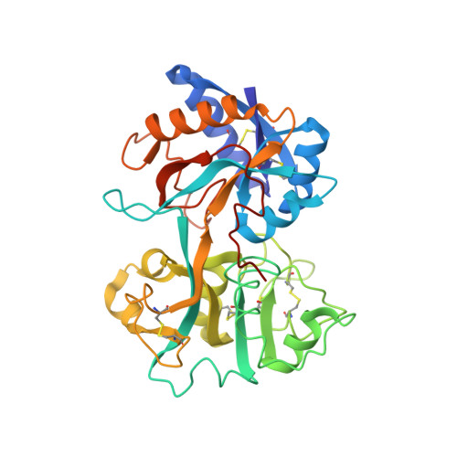
    A conserved arginine residue helps to form the synergistic anion binding site in transferrins. To probe the importance of this residue for anion binding and iron binding, Arg 121 has been mutated to Ser and Glu in N-terminal half-molecule of human lactoferrin. The two mutants, R121S and R121E, have been expressed, purified, and crystallized. Their three-dimensional structures have been determined by X-ray diffraction at 2.3 and 2.5 A resolution, respectively. The structures were determined by molecular replacement and were refined by restrained least squares methods to final R values of 0.185 and 0.204. Both mutants still bind iron but with decreased stability. The crystal structures show that destabilization of iron binding probably results from disruption of the anion binding site; mutation of Arg 121 removes one wall of the anion binding pocket and causes the synergistic carbonate ion to be displaced 0.5 A from its position in the wild-type protein. In the process it becomes partially detached from the helix N-terminus that forms the rest of the anion binding site.


  • Organizational Affiliation

    Department of Biochemistry, Massey University, Palmerston North, New Zealand.


Macromolecules
Find similar proteins by:  (by identity cutoff)  |  3D Structure
Entity ID: 1
MoleculeChains Sequence LengthOrganismDetailsImage
HUMAN LACTOFERRIN333Homo sapiensMutation(s): 2 
Gene Names: LFN
UniProt & NIH Common Fund Data Resources
Find proteins for P02788 (Homo sapiens)
Explore P02788 
Go to UniProtKB:  P02788
PHAROS:  P02788
GTEx:  ENSG00000012223 
Entity Groups  
Sequence Clusters30% Identity50% Identity70% Identity90% Identity95% Identity100% Identity
UniProt GroupP02788
Sequence Annotations
Expand
  • Reference Sequence
Experimental Data & Validation

Experimental Data

  • Method: X-RAY DIFFRACTION
  • Resolution: 2.30 Å
  • R-Value Free: 0.270 
  • R-Value Work: 0.196 
  • Space Group: C 1 2 1
Unit Cell:
Length ( Å )Angle ( ˚ )
a = 97.9α = 90
b = 78.8β = 99.2
c = 58.9γ = 90
Software Package:
Software NamePurpose
RIGAKUdata collection
AGROVATA/CCP4data reduction
CCP4model building
TNTrefinement
RIGAKUdata reduction
Agrovatadata scaling
CCP4phasing

Structure Validation

View Full Validation Report


Entry History 

Deposition Data

Revision History  (Full details and data files)

  • Version 1.0: 1997-04-01
    Type: Initial release
  • Version 1.1: 2008-03-24
    Changes: Version format compliance
  • Version 1.2: 2011-07-13
    Changes: Version format compliance
  • Version 1.3: 2017-11-29
    Changes: Derived calculations, Other, Refinement description
  • Version 1.4: 2021-11-03
    Changes: Database references, Derived calculations
  • Version 1.5: 2023-08-09
    Changes: Refinement description