1V6X

Crystal Structure Of Xylanase From Streptomyces Olivaceoviridis E-86 Complexed With 3(3)-4-O-methyl-alpha-D-glucuronosyl-xylotriose


Experimental Data Snapshot

  • Method: X-RAY DIFFRACTION
  • Resolution: 2.10 Å
  • R-Value Free: 0.235 
  • R-Value Work: 0.194 
  • R-Value Observed: 0.194 

wwPDB Validation   3D Report Full Report


This is version 2.1 of the entry. See complete history


Literature

Crystal structures of decorated xylooligosaccharides bound to a family 10 xylanase from Streptomyces olivaceoviridis E-86

Fujimoto, Z.Kaneko, S.Kuno, A.Kobayashi, H.Kusakabe, I.Mizuno, H.

(2004) J Biol Chem 279: 9606-9614

  • DOI: https://doi.org/10.1074/jbc.M312293200
  • Primary Citation of Related Structures:  
    1V6U, 1V6V, 1V6W, 1V6X

  • PubMed Abstract: 

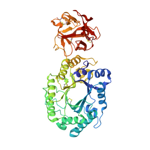
    The family 10 xylanase from Streptomyces olivaceoviridis E-86 (SoXyn10A) consists of a GH10 catalytic domain, which is joined by a Gly/Pro-rich linker to a family 13 carbohydrate-binding module (CBM13) that interacts with xylan. To understand how GH10 xylanases and CBM13 recognize decorated xylans, the crystal structure of SoXyn10A was determined in complex with alpha-l-arabinofuranosyl- and 4-O-methyl-alpha-d-glucuronosyl-xylooligosaccharides. The bound sugars were observed in the subsites of the catalytic cleft and also in subdomains alpha and gamma of CBM13. The data reveal that the binding mode of the oligosaccharides in the active site of the catalytic domain is entirely consistent with the substrate specificity and, in conjunction with the accompanying paper, demonstrate that the accommodation of the side chains in decorated xylans is conserved in GH10 xylanases of SoXyn10A against arabinoglucuronoxylan. CBM13 was shown to bind xylose or xylooligosaccharides reversibly by using nonsymmetric sugars as the ligands. The independent multiple sites in CBM13 may increase the probability of substrate binding.


  • Organizational Affiliation

    Department of Biochemistry, National Institute of Agrobiological Sciences, Tsukuba, Ibaraki 305-8602, Japan. zui@affrc.go.jp


Macromolecules
Find similar proteins by:  (by identity cutoff)  |  3D Structure
Entity ID: 1
MoleculeChains Sequence LengthOrganismDetailsImage
ENDO-1,4-BETA-D-XYLANASE
A, B
436Streptomyces olivaceoviridisMutation(s): 0 
EC: 3.2.1.8
UniProt
Find proteins for Q7SI98 (Streptomyces olivaceoviridis)
Explore Q7SI98 
Go to UniProtKB:  Q7SI98
Entity Groups  
Sequence Clusters30% Identity50% Identity70% Identity90% Identity95% Identity100% Identity
UniProt GroupQ7SI98
Sequence Annotations
Expand
  • Reference Sequence
Oligosaccharides

Help

Entity ID: 2
MoleculeChains Length2D Diagram Glycosylation3D Interactions
4-O-methyl-alpha-D-glucopyranuronic acid-(1-2)-beta-D-xylopyranose-(1-4)-beta-D-xylopyranose-(1-4)-beta-D-xylopyranose
C, E, G
4N/A
Glycosylation Resources
GlyTouCan:  G75464GU
GlyCosmos:  G75464GU
Entity ID: 3
MoleculeChains Length2D Diagram Glycosylation3D Interactions
beta-D-xylopyranose-(1-4)-beta-D-xylopyranose
D, H
2N/A
Glycosylation Resources
GlyTouCan:  G87728WL
GlyCosmos:  G87728WL
Entity ID: 4
MoleculeChains Length2D Diagram Glycosylation3D Interactions
beta-D-xylopyranose-(1-4)-beta-D-xylopyranose-(1-4)-beta-D-xylopyranose
F
3N/A
Glycosylation Resources
GlyTouCan:  G87429QT
GlyCosmos:  G87429QT
Small Molecules
Ligands 1 Unique
IDChains Name / Formula / InChI Key2D Diagram3D Interactions
XYP
Query on XYP

Download Ideal Coordinates CCD File 
I [auth B],
J [auth B]
beta-D-xylopyranose
C5 H10 O5
SRBFZHDQGSBBOR-KKQCNMDGSA-N
Biologically Interesting Molecules (External Reference) 2 Unique
Experimental Data & Validation

Experimental Data

  • Method: X-RAY DIFFRACTION
  • Resolution: 2.10 Å
  • R-Value Free: 0.235 
  • R-Value Work: 0.194 
  • R-Value Observed: 0.194 
  • Space Group: P 21 21 21
Unit Cell:
Length ( Å )Angle ( ˚ )
a = 74.54α = 90
b = 94.24β = 90
c = 137.43γ = 90
Software Package:
Software NamePurpose
CNSrefinement
CrystalCleardata reduction
CrystalCleardata scaling
CNSphasing

Structure Validation

View Full Validation Report


Entry History 

Deposition Data

Revision History  (Full details and data files)

  • Version 1.0: 2004-04-27
    Type: Initial release
  • Version 1.1: 2008-04-27
    Changes: Version format compliance
  • Version 1.2: 2011-07-13
    Changes: Version format compliance
  • Version 2.0: 2020-07-29
    Type: Remediation
    Reason: Carbohydrate remediation
    Changes: Atomic model, Data collection, Derived calculations, Structure summary
  • Version 2.1: 2023-10-25
    Changes: Data collection, Database references, Derived calculations, Refinement description, Structure summary