1UZJ | pdb_00001uzj

Integrin binding cbEGF22-TB4-cbEGF33 fragment of human fibrillin-1, holo form.


Experimental Data Snapshot

  • Method: X-RAY DIFFRACTION
  • Resolution: 2.25 Å
  • R-Value Free: 
    0.318 (Depositor) 
  • R-Value Work: 
    0.247 (Depositor) 
  • R-Value Observed: 
    0.247 (Depositor) 

Starting Model: other
View more details

wwPDB Validation 3D Report Full Report

Validation slider image for 1UZJ

This is version 1.4 of the entry. See complete history

Literature

Structure of the Integrin Binding Fragment from Fibrillin-1 Gives New Insights Into Microfibril Organization

Lee, S.S.J.Knott, V.Jovanovi, J.Harlos, K.Grimes, J.Choulier, L.Mardon, H.Stuart, D.I.Handford, P.A.

(2004) Structure 12: 717

  • DOI: https://doi.org/10.1016/j.str.2004.02.023
  • Primary Citation Related Structures: 
    1UZJ, 1UZK, 1UZP, 1UZQ

  • PubMed Abstract: 

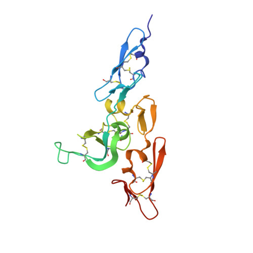
    Human fibrillin-1, the major structural protein of extracellular matrix (ECM) 10-12 nm microfibrils, is dominated by 43 calcium binding epidermal growth factor-like (cbEGF) and 7 transforming growth factor beta binding protein-like (TB) domains. Crystal structures reveal the integrin binding cbEGF22-TB4-cbEGF23 fragment of human fibrillin-1 to be a Ca(2+)-rigidified tetragonal pyramid. We suggest that other cbEGF-TB pairs within the fibrillins may adopt a similar orientation to cbEGF22-TB4. In addition, we have located a flexible RGD integrin binding loop within TB4. Modeling, cell attachment and spreading assays, immunocytochemistry, and surface plasmon resonance indicate that cbEGF22 bound to TB4 is a requirement for integrin activation and provide insight into the molecular basis of the fibrillin-1 interaction with alphaVbeta3. In light of our data, we propose a novel model for the assembly of the fibrillin microfibril and a mechanism to explain its extensibility.


  • Organizational Affiliation
    • The Henry Wellcome Building of Genomic Medicine, Roosevelt Drive, Oxford OX3 7BN, United Kingdom.

Macromolecule Content 

  • Total Structure Weight: 52.24 kDa 
  • Atom Count: 3,806 
  • Modeled Residue Count: 486 
  • Deposited Residue Count: 486 
  • Unique protein chains: 1

Macromolecules

Find similar proteins by:|  3D Structure
Entity ID: 1
MoleculeChains  Sequence LengthOrganismDetailsImage
FIBRILLIN-1
A, B, C
162Homo sapiensMutation(s): 0 
UniProt & NIH Common Fund Data Resources
Find proteins for P35555 (Homo sapiens)
Explore P35555 
Go to UniProtKB:  P35555
PHAROS:  P35555
GTEx:  ENSG00000166147 
Entity Groups
Sequence Clusters30% Identity50% Identity70% Identity90% Identity95% Identity100% Identity
UniProt GroupP35555
Sequence Annotations
Expand
Reference Sequence

Experimental Data & Validation

Experimental Data

  • Method: X-RAY DIFFRACTION
  • Resolution: 2.25 Å
  • R-Value Free:  0.318 (Depositor) 
  • R-Value Work:  0.247 (Depositor) 
  • R-Value Observed: 0.247 (Depositor) 
Space Group: P 1 21 1
Unit Cell:
Length ( Å )Angle ( ˚ )
a = 43.1α = 90
b = 105.6β = 104.2
c = 65γ = 90
Software Package:
Software NamePurpose
CNSrefinement
DENZOdata reduction
SCALEPACKdata scaling
CNSphasing

Structure Validation

View Full Validation Report


Entry History 

Deposition Data

Revision History  (Full details and data files)

  • Version 1.0: 2004-04-08
    Type: Initial release
  • Version 1.1: 2011-05-07
    Changes: Version format compliance
  • Version 1.2: 2011-07-13
    Changes: Version format compliance
  • Version 1.3: 2024-05-01
    Changes: Data collection, Database references, Derived calculations, Refinement description
  • Version 1.4: 2024-10-16
    Changes: Structure summary