1UYE | pdb_00001uye

Human Hsp90-alpha with 8-(2-chloro-3,4,5-trimethoxy-benzyl)-9-pent-4-ylnyl-9H-purin-6-ylamine


Experimental Data Snapshot

  • Method: X-RAY DIFFRACTION
  • Resolution: 2.00 Å
  • R-Value Free: 
    0.218 (Depositor), 0.247 (DCC) 
  • R-Value Work: 
    0.183 (Depositor), 0.195 (DCC) 

Starting Model: experimental
View more details

wwPDB Validation 3D Report Full Report

Validation slider image for 1UYE

Ligand Structure Quality Assessment 


This is version 1.4 of the entry. See complete history

Literature

Structure-Activity Relationships in Purine-Based Inhibitor Binding to Hsp90 Isoforms

Wright, L.Barril, X.Dymock, B.Sheridan, L.Surgenor, A.Beswick, M.Drysdale, M.Collier, A.Massey, A.Davies, N.Fink, A.Fromont, C.Aherne, W.Boxall, K.Sharp, S.Workman, P.Hubbard, R.E.

(2004) Chem Biol 11: 775

  • DOI: https://doi.org/10.1016/j.chembiol.2004.03.033
  • Primary Citation Related Structures: 
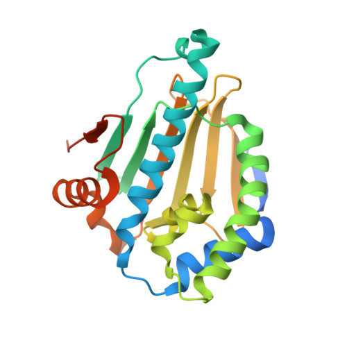
    1UY6, 1UY7, 1UY8, 1UY9, 1UYC, 1UYD, 1UYE, 1UYF, 1UYG, 1UYH, 1UYI, 1UYK, 1UYL, 1UYM

  • PubMed Abstract: 

    Inhibition of the ATPase activity of the chaperone protein HSP90 is a potential strategy for treatment of cancers. We have determined structures of the HSP90alpha N-terminal domain complexed with the purine-based inhibitor, PU3, and analogs with enhanced potency both in enzyme and cell-based assays. The compounds induce upregulation of HSP70 and downregulation of the known HSP90 client proteins Raf-1, CDK4, and ErbB2, confirming that the molecules inhibit cell growth by a mechanism dependent on HSP90 inhibition. We have also determined the first structure of the N-terminal domain of HSP90beta, complexed with PU3. The structures allow a detailed rationale to be developed for the observed affinity of the PU3 class of compounds for HSP90 and also provide a structural framework for design of compounds with improved binding affinity and drug-like properties.


  • Organizational Affiliation
    • Vernalis (R&D) Ltd., Granta Park, Abington, Cambridge CB1 6GB, UK.

Macromolecule Content 

  • Total Structure Weight: 27.02 kDa 
  • Atom Count: 1,951 
  • Modeled Residue Count: 209 
  • Deposited Residue Count: 236 
  • Unique protein chains: 1

Macromolecules

Find similar proteins by:|  3D Structure
Entity ID: 1
MoleculeChains  Sequence LengthOrganismDetailsImage
HEAT SHOCK PROTEIN HSP 90-ALPHA236Homo sapiensMutation(s): 0 
EC: 3.6.4.10
UniProt & NIH Common Fund Data Resources
Find proteins for P07900 (Homo sapiens)
Explore P07900 
Go to UniProtKB:  P07900
PHAROS:  P07900
GTEx:  ENSG00000080824 
Entity Groups
Sequence Clusters30% Identity50% Identity70% Identity90% Identity95% Identity100% Identity
UniProt GroupP07900
Sequence Annotations
Expand
Reference Sequence

Small Molecules

Ligands 1 Unique
IDChains Name / Formula / InChI Key2D Diagram3D Interactions
PU9

Query on PU9


Download:Ideal Coordinates CCD File
B [auth A]8-(2-CHLORO-3,4,5-TRIMETHOXY-BENZYL)-9-PENT-4-YLNYL-9H-PURIN-6-YLAMINE
C20 H22 Cl N5 O3
WUJFGZAAFADPSF-UHFFFAOYSA-N
Binding Affinity Annotations 
IDSourceBinding Affinity
PU9 BindingDB:  1UYE IC50: 2.00e+5 (nM) from 1 assay(s)

Experimental Data & Validation

Experimental Data

  • Method: X-RAY DIFFRACTION
  • Resolution: 2.00 Å
  • R-Value Free:  0.218 (Depositor), 0.247 (DCC) 
  • R-Value Work:  0.183 (Depositor), 0.195 (DCC) 
Space Group: I 2 2 2
Unit Cell:
Length ( Å )Angle ( ˚ )
a = 66.7α = 90
b = 90.755β = 90
c = 98.171γ = 90
Software Package:
Software NamePurpose
REFMACrefinement
d*TREKdata reduction
d*TREKdata scaling
MOLREPphasing

Structure Validation

View Full Validation Report


Ligand Structure Quality Assessment 


Entry History 

Revision History  (Full details and data files)

  • Version 1.0: 2004-07-01
    Type: Initial release
  • Version 1.1: 2011-05-07
    Changes: Version format compliance
  • Version 1.2: 2011-07-13
    Changes: Version format compliance
  • Version 1.3: 2017-07-05
    Changes: Data collection
  • Version 1.4: 2023-12-13
    Changes: Data collection, Database references, Other, Refinement description