A Closed Conformation of Bacillus Subtilis Oxalate Decarboxylase Oxdc Provides Evidence for the True Identity of the Active Site
Just, V.J., Stevenson, C.E.M., Bowater, L., Tanner, A., Lawson, D.M., Bornemann, S.(2004) J Biological Chem 279: 19867
- PubMed: 14871895 Search on PubMed
- DOI: https://doi.org/10.1074/jbc.M313820200
- Primary Citation Related Structures:
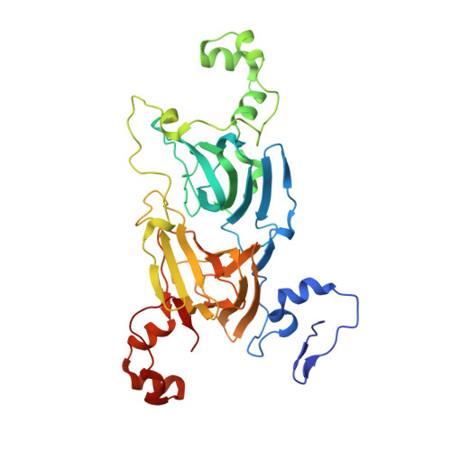
1UW8 - PubMed Abstract:
Oxalate decarboxylase (EC 4.1.1.2) catalyzes the conversion of oxalate to formate and carbon dioxide and utilizes dioxygen as a cofactor. By contrast, the evolutionarily related oxalate oxidase (EC 1.2.3.4) converts oxalate and dioxygen to carbon dioxide and hydrogen peroxide. Divergent free radical catalytic mechanisms have been proposed for these enzymes that involve the requirement of an active site proton donor in the decarboxylase but not the oxidase reaction. The oxidase possesses only one domain and manganese binding site per subunit, while the decarboxylase has two domains and two manganese sites per subunit. A structure of the decarboxylase together with a limited mutagenesis study has recently been interpreted as evidence that the C-terminal domain manganese binding site (site 2) is the catalytic site and that Glu-333 is the crucial proton donor (Anand, R., Dorrestein, P. C., Kinsland, C., Begley, T. P., and Ealick, S. E. (2002) Biochemistry 41, 7659-7669). The N-terminal binding site (site 1) of this structure is solvent-exposed (open) and lacks a suitable proton donor for the decarboxylase reaction. We report a new structure of the decarboxylase that shows a loop containing a 3(10) helix near site 1 in an alternative conformation. This loop adopts a "closed" conformation forming a lid covering the entrance to site 1. This conformational change brings Glu-162 close to the manganese ion, making it a new candidate for the crucial proton donor. Site-directed mutagenesis of equivalent residues in each domain provides evidence that Glu-162 performs this vital role and that the N-terminal domain is either the sole or the dominant catalytically active domain.
- Biological Chemistry Department, John Innes Centre, Norwich Research Park, Colney, Norwich NR4 7UH, United Kingdom.
Organizational Affiliation: