1US1

Crystal structure of human vascular adhesion protein-1


Experimental Data Snapshot

  • Method: X-RAY DIFFRACTION
  • Resolution: 2.90 Å
  • R-Value Free: 0.267 
  • R-Value Work: 0.241 
  • R-Value Observed: 0.242 

wwPDB Validation   3D Report Full Report


Ligand Structure Quality Assessment 


This is version 2.1 of the entry. See complete history


Literature

Crystal Structure of the Human Vascular Adhesion Protein-1: Unique Structural Features with Functional Implications.

Airenne, T.T.Nymalm, Y.Kidron, H.Smith, D.J.Pihlavisto, M.Salmi, M.Jalkanen, S.Johnson, M.S.Salminen, T.A.

(2005) Protein Sci 14: 1964

  • DOI: https://doi.org/10.1110/ps.051438105
  • Primary Citation of Related Structures:  
    1PU4, 1US1

  • PubMed Abstract: 

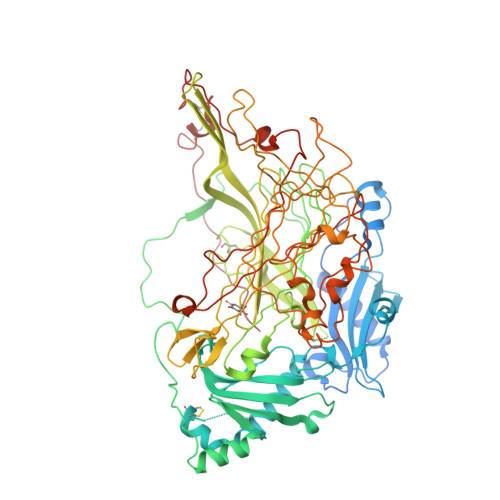
    The expression of human vascular adhesion protein-1 (hVAP-1) is induced at sites of inflammation where extravasation of lymphocytes from blood to the peripheral tissue occurs. We have solved the X-ray structure of hVAP-1, a human copper amine oxidase (CAO), which is distinguished from other CAOs in being membrane-bound. The dimer structure reveals some intriguing features that may have fundamental roles in the adhesive and enzymatic functions of hVAP-1, especially regarding the role of hVAP-1 in inflammation, lymphocyte attachment, and signaling. Firstly, Leu469 at the substrate channel may play a key role in controlling the substrate entry; depending on its conformation, it either blocks or gives access to the active site. Secondly, sugar units are clearly observed at two of the six predicted N-glycosylation sites. Moreover, mutagenesis analysis showed that all of the predicted sites were glycosylated in the protein used for crystallization. Thirdly, the existence of a solvent-exposed RGD motif at the entrance to each active site in hVAP-1 suggests that it may have a functional role.


  • Organizational Affiliation

    Department of Biochemistry and Pharmacy, Abo Akademi University, FIN-20520 Turku, Finland.


Macromolecules
Find similar proteins by:  (by identity cutoff)  |  3D Structure
Entity ID: 1
MoleculeChains Sequence LengthOrganismDetailsImage
MEMBRANE COPPER AMINE OXIDASE
A, B
763Homo sapiensMutation(s): 0 
EC: 1.4.3.6
UniProt & NIH Common Fund Data Resources
Find proteins for Q16853 (Homo sapiens)
Explore Q16853 
Go to UniProtKB:  Q16853
PHAROS:  Q16853
GTEx:  ENSG00000131471 
Entity Groups  
Sequence Clusters30% Identity50% Identity70% Identity90% Identity95% Identity100% Identity
UniProt GroupQ16853
Sequence Annotations
Expand
  • Reference Sequence
Oligosaccharides

Help

Entity ID: 2
MoleculeChains Length2D Diagram Glycosylation3D Interactions
2-acetamido-2-deoxy-beta-D-glucopyranose-(1-4)-2-acetamido-2-deoxy-beta-D-glucopyranose
C, D
2N-Glycosylation
Glycosylation Resources
GlyTouCan:  G42666HT
GlyCosmos:  G42666HT
GlyGen:  G42666HT
Small Molecules
Ligands 3 Unique
IDChains Name / Formula / InChI Key2D Diagram3D Interactions
NAG
Query on NAG

Download Ideal Coordinates CCD File 
H [auth A],
L [auth B]
2-acetamido-2-deoxy-beta-D-glucopyranose
C8 H15 N O6
OVRNDRQMDRJTHS-FMDGEEDCSA-N
CU
Query on CU

Download Ideal Coordinates CCD File 
E [auth A],
I [auth B]
COPPER (II) ION
Cu
JPVYNHNXODAKFH-UHFFFAOYSA-N
CA
Query on CA

Download Ideal Coordinates CCD File 
F [auth A],
G [auth A],
J [auth B],
K [auth B]
CALCIUM ION
Ca
BHPQYMZQTOCNFJ-UHFFFAOYSA-N
Modified Residues  1 Unique
IDChains TypeFormula2D DiagramParent
TPQ
Query on TPQ
A, B
L-PEPTIDE LINKINGC9 H9 N O5TYR
Experimental Data & Validation

Experimental Data

  • Method: X-RAY DIFFRACTION
  • Resolution: 2.90 Å
  • R-Value Free: 0.267 
  • R-Value Work: 0.241 
  • R-Value Observed: 0.242 
  • Space Group: P 65 2 2
Unit Cell:
Length ( Å )Angle ( ˚ )
a = 226.099α = 90
b = 226.099β = 90
c = 223.004γ = 120
Software Package:
Software NamePurpose
REFMACrefinement
XDSdata reduction
XDSdata scaling

Structure Validation

View Full Validation Report


Ligand Structure Quality Assessment 


Entry History 

Deposition Data

Revision History  (Full details and data files)

  • Version 1.0: 2005-02-16
    Type: Initial release
  • Version 1.1: 2011-05-08
    Changes: Version format compliance
  • Version 1.2: 2011-07-13
    Changes: Version format compliance
  • Version 1.3: 2018-02-28
    Changes: Data collection
  • Version 2.0: 2020-07-29
    Type: Remediation
    Reason: Carbohydrate remediation
    Changes: Atomic model, Data collection, Derived calculations, Other, Structure summary
  • Version 2.1: 2023-12-13
    Changes: Data collection, Database references, Refinement description, Structure summary