1UOG

Deacetoxycephalosporin C synthase complexed with deacetoxycephalosporin C


Experimental Data Snapshot

  • Method: X-RAY DIFFRACTION
  • Resolution: 1.70 Å
  • R-Value Free: 0.242 
  • R-Value Work: 0.204 
  • R-Value Observed: 0.206 

wwPDB Validation   3D Report Full Report


Ligand Structure Quality Assessment 


This is version 1.4 of the entry. See complete history


Literature

The Structural Basis of Cephalosporin Formation in a Mononuclear Ferrous Enzyme

Valegard, K.Terwisscha van Scheltinga, A.C.Dubus, A.Ranghino, G.Oster, L.M.Hajdu, J.Andersson, I.

(2004) Nat Struct Mol Biol 11: 95-101

  • DOI: https://doi.org/10.1038/nsmb712
  • Primary Citation of Related Structures:  
    1UNB, 1UO9, 1UOB, 1UOF, 1UOG

  • PubMed Abstract: 

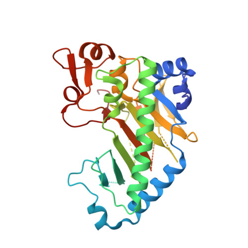
    Deacetoxycephalosporin-C synthase (DAOCS) is a mononuclear ferrous enzyme that transforms penicillins into cephalosporins by inserting a carbon atom into the penicillin nucleus. In the first half-reaction, dioxygen and 2-oxoglutarate produce a reactive iron-oxygen species, succinate and CO2. The oxidizing iron species subsequently reacts with penicillin to give cephalosporin and water. Here we describe high-resolution structures for ferrous DAOCS in complex with penicillins, the cephalosporin product, the cosubstrate and the coproduct. Steady-state kinetic data, quantum-chemical calculations and the new structures indicate a reaction sequence in which a 'booby-trapped' oxidizing species is formed. This species is stabilized by the negative charge of succinate on the iron. The binding sites of succinate and penicillin overlap, and when penicillin replaces succinate, it removes the stabilizing charge, eliciting oxidative attack on itself. Requisite groups of penicillin are within 1 A of the expected position of a ferryl oxygen in the enzyme-penicillin complex.


  • Organizational Affiliation

    Molecular Biophysics, Department of Cellular and Molecular Biology, Uppsala University, Box 596, S-751 24 Uppsala, Sweden.


Macromolecules
Find similar proteins by:  (by identity cutoff)  |  3D Structure
Entity ID: 1
MoleculeChains Sequence LengthOrganismDetailsImage
DEACETOXYCEPHALOSPORIN C SYNTHETASE311Streptomyces clavuligerusMutation(s): 0 
EC: 1.14.20.1
UniProt
Find proteins for P18548 (Streptomyces clavuligerus)
Explore P18548 
Go to UniProtKB:  P18548
Entity Groups  
Sequence Clusters30% Identity50% Identity70% Identity90% Identity95% Identity100% Identity
UniProt GroupP18548
Sequence Annotations
Expand
  • Reference Sequence
Small Molecules
Ligands 2 Unique
IDChains Name / Formula / InChI Key2D Diagram3D Interactions
P1C
Query on P1C

Download Ideal Coordinates CCD File 
B [auth A]DEACETOXYCEPHALOSPORIN-C
C14 H19 N3 O6 S
NNQIJOYQWYKBOW-JWKOBGCHSA-N
FE2
Query on FE2

Download Ideal Coordinates CCD File 
C [auth A]FE (II) ION
Fe
CWYNVVGOOAEACU-UHFFFAOYSA-N
Experimental Data & Validation

Experimental Data

  • Method: X-RAY DIFFRACTION
  • Resolution: 1.70 Å
  • R-Value Free: 0.242 
  • R-Value Work: 0.204 
  • R-Value Observed: 0.206 
  • Space Group: H 3
Unit Cell:
Length ( Å )Angle ( ˚ )
a = 106.6α = 90
b = 106.6β = 90
c = 74γ = 120
Software Package:
Software NamePurpose
REFMACrefinement
DENZOdata reduction
SCALEPACKdata scaling

Structure Validation

View Full Validation Report


Ligand Structure Quality Assessment 


Entry History 

Deposition Data

Revision History  (Full details and data files)

  • Version 1.0: 2004-01-09
    Type: Initial release
  • Version 1.1: 2011-05-08
    Changes: Version format compliance
  • Version 1.2: 2011-07-13
    Changes: Version format compliance
  • Version 1.3: 2018-01-17
    Changes: Data collection, Database references, Structure summary
  • Version 1.4: 2023-12-13
    Changes: Data collection, Database references, Derived calculations, Other, Refinement description