1UKF | pdb_00001ukf

Crystal Structure of Pseudomonas Avirulence Protein AvrPphB


Experimental Data Snapshot

  • Method: X-RAY DIFFRACTION
  • Resolution: 1.35 Å
  • R-Value Free: 
    0.222 (Depositor), 0.219 (DCC) 
  • R-Value Work: 
    0.204 (Depositor), 0.202 (DCC) 
  • R-Value Observed: 
    0.204 (Depositor) 

wwPDB Validation 3D Report Full Report

Validation slider image for 1UKF

This is version 1.3 of the entry. See complete history

Literature

The crystal structure of Pseudomonas avirulence protein AvrPphB: a papain-like fold with a distinct substrate-binding site.

Zhu, M.Shao, F.Innes, R.W.Dixon, J.E.Xu, Z.

(2004) Proc Natl Acad Sci U S A 101: 302-307

  • DOI: https://doi.org/10.1073/pnas.2036536100
  • Primary Citation Related Structures: 
    1UKF

  • PubMed Abstract: 

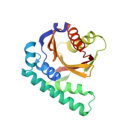
    AvrPphB is an avirulence (Avr) protein from the plant pathogen Pseudomonas syringae that can trigger a disease-resistance response in a number of host plants including Arabidopsis. AvrPphB belongs to a novel family of cysteine proteases with the charter member of this family being the Yersinia effector protein YopT. AvrPphB has a very stringent substrate specificity, catalyzing a single proteolytic cleavage in the Arabidopsis serine/threonine kinase PBS1. We have determined the crystal structure of AvrPphB by x-ray crystallography at 1.35-A resolution. The structure is composed of a central antiparallel beta-sheet, with alpha-helices packing on both sides of the sheet to form a two-lobe structure. The core of this structure resembles the papain-like cysteine proteases. The similarity includes the AvrPphB active site catalytic triad of Cys-98, His-212, and Asp-227 and the oxyanion hole residue Asn-93. Based on analogy with inhibitor complexes of the papain-like proteases, we propose a model for the substrate-binding mechanism of AvrPphB. A deep and positively charged pocket (S2) and a neighboring shallow surface (S3) likely bind to aspartic acid and glycine residues in the substrate located two (P2) and three (P3) residues N terminal to the cleavage site, respectively. Further implications about the specificity of plant pathogen recognition are also discussed.


  • Organizational Affiliation
    • Department of Biological Chemistry, Medical School and Life Sciences Institute, University of Michigan, Ann Arbor, MI 48109, USA.

Macromolecule Content 

  • Total Structure Weight: 20.39 kDa 
  • Atom Count: 1,605 
  • Modeled Residue Count: 188 
  • Deposited Residue Count: 188 
  • Unique protein chains: 1

Macromolecules

Find similar proteins by:|  3D Structure
Entity ID: 1
MoleculeChains  Sequence LengthOrganismDetailsImage
Avirulence protein AVRPPH3188Pseudomonas savastanoi pv. phaseolicolaMutation(s): 0 
EC: 3.4.22
UniProt
Find proteins for Q52430 (Pseudomonas savastanoi pv. phaseolicola)
Explore Q52430 
Go to UniProtKB:  Q52430
Entity Groups
Sequence Clusters30% Identity50% Identity70% Identity90% Identity95% Identity100% Identity
UniProt GroupQ52430
Sequence Annotations
Expand
Reference Sequence

Experimental Data & Validation

Experimental Data

  • Method: X-RAY DIFFRACTION
  • Resolution: 1.35 Å
  • R-Value Free:  0.222 (Depositor), 0.219 (DCC) 
  • R-Value Work:  0.204 (Depositor), 0.202 (DCC) 
  • R-Value Observed: 0.204 (Depositor) 
Space Group: P 21 21 21
Unit Cell:
Length ( Å )Angle ( ˚ )
a = 43.601α = 90
b = 49.596β = 90
c = 75.705γ = 90
Software Package:
Software NamePurpose
CNSrefinement
HKL-2000data reduction
SCALEPACKdata scaling
CNSphasing

Structure Validation

View Full Validation Report


Entry History 

Deposition Data

Revision History  (Full details and data files)

  • Version 1.0: 2003-12-09
    Type: Initial release
  • Version 1.1: 2008-04-27
    Changes: Version format compliance
  • Version 1.2: 2011-07-13
    Changes: Version format compliance
  • Version 1.3: 2023-12-27
    Changes: Data collection, Database references