1UBZ

Crystal structure of Glu102-mutant human lysozyme doubly labeled with 2',3'-epoxypropyl beta-glycoside of N-acetyllactosamine


Experimental Data Snapshot

  • Method: X-RAY DIFFRACTION
  • Resolution: 2.00 Å
  • R-Value Free: 0.266 
  • R-Value Work: 0.189 

wwPDB Validation   3D Report Full Report


This is version 2.2 of the entry. See complete history


Literature

X-ray structural analysis of the ligand-recognition mechanism in the dual-affinity labeling of c-type lysozyme with 2',3'-epoxypropyl beta-glycoside of N-acetyllactosamine

Muraki, M.Harata, K.

(2003) J Mol Recognit 16: 72-82

  • DOI: https://doi.org/10.1002/jmr.611
  • Primary Citation of Related Structures:  
    1UBZ, 1UC0

  • PubMed Abstract: 

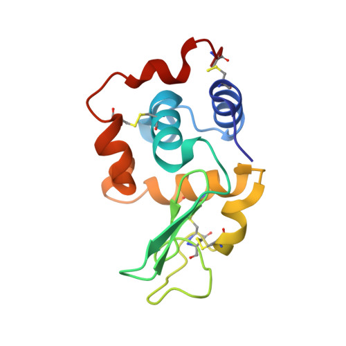
    In spite of the belonging to the same c-type lysozyme family, hen egg-white lysozyme (HEWL) was much less susceptible to the dual-affinity labeling with 2',3'-epoxypropyl beta-glycoside of N-acetyllactosamine (Galbeta1,4GlcNAc-Epo) than human lysozyme (HL). The three-dimensional structures of the HEWL labeled with single Galbeta1,4GlcNAc-Epo and the Glu102-mutant HL labeled with double Galbeta1,4GlcNAc-Epo were determined by X-ray crystallography at resolutions of 1.85 and 2.0 A, respectively. The overall conformation and the interaction mode of the carbohydrate ligand part in the singly labeled HEWL and the doubly labeled Glu102-mutant HL were basically identical to those of the correspondingly labeled wild-type HL with minor alterations in some stereochemical parameters. A detailed comparison of the structures revealed the key protein-carbohydrate and carbohydrate-carbohydrate interactions essential for the dual labeling. It was suggested that the difference in the efficiency of the dual labeling was caused by the structural difference between Gln104 in HL and Asn103 in HEWL. The relevance to our previous study and the carbohydrate-carbohydrate interaction on cell-surface membranes were discussed.


  • Organizational Affiliation

    Biological Information Research Center, National Institute of Advanced Industrial Science and Technology, Tsukuba, Ibaraki 305-8566, Japan. m-muraki@aist.go.jp


Macromolecules
Find similar proteins by:  (by identity cutoff)  |  3D Structure
Entity ID: 1
MoleculeChains Sequence LengthOrganismDetailsImage
Lysozyme C130Homo sapiensMutation(s): 1 
EC: 3.2.1.17
UniProt & NIH Common Fund Data Resources
Find proteins for P61626 (Homo sapiens)
Explore P61626 
Go to UniProtKB:  P61626
PHAROS:  P61626
GTEx:  ENSG00000090382 
Entity Groups  
Sequence Clusters30% Identity50% Identity70% Identity90% Identity95% Identity100% Identity
UniProt GroupP61626
Sequence Annotations
Expand
  • Reference Sequence
Oligosaccharides

Help

Entity ID: 2
MoleculeChains Length2D Diagram Glycosylation3D Interactions
beta-D-galactopyranose-(1-4)-2-acetamido-2-deoxy-beta-D-glucopyranose
B, C
2N/A
Glycosylation Resources
GlyTouCan:  G00055MO
GlyCosmos:  G00055MO
GlyGen:  G00055MO
Experimental Data & Validation

Experimental Data

  • Method: X-RAY DIFFRACTION
  • Resolution: 2.00 Å
  • R-Value Free: 0.266 
  • R-Value Work: 0.189 
  • Space Group: P 21 21 21
Unit Cell:
Length ( Å )Angle ( ˚ )
a = 56.33α = 90
b = 61β = 90
c = 32.71γ = 90
Software Package:
Software NamePurpose
MOSFLMdata reduction
TRUNCATEdata reduction
X-PLORmodel building
X-PLORrefinement
CCP4data scaling
X-PLORphasing

Structure Validation

View Full Validation Report


Entry History 

Deposition Data

Revision History  (Full details and data files)

  • Version 1.0: 2003-04-22
    Type: Initial release
  • Version 1.1: 2008-04-27
    Changes: Version format compliance
  • Version 1.2: 2011-07-13
    Changes: Non-polymer description, Version format compliance
  • Version 2.0: 2020-07-29
    Type: Remediation
    Reason: Carbohydrate remediation
    Changes: Atomic model, Data collection, Derived calculations, Structure summary
  • Version 2.1: 2021-11-10
    Changes: Database references, Structure summary
  • Version 2.2: 2023-12-27
    Changes: Data collection