1U68 | pdb_00001u68

DHNA 7,8 DIHYDRONEOPTERIN COMPLEX


Experimental Data Snapshot

  • Method: X-RAY DIFFRACTION
  • Resolution: 2.40 Å
  • R-Value Free: 
    0.368 (Depositor) 
  • R-Value Work: 
    0.279 (Depositor) 

wwPDB Validation 3D Report Full Report

Validation slider image for 1U68

This is version 1.3 of the entry. See complete history

Literature

Discovery of potent inhibitors of dihydroneopterin aldolase using CrystaLEAD high-throughput X-ray crystallographic screening and structure-directed lead optimization.

Sanders, W.J.Nienaber, V.L.Lerner, C.G.McCall, J.O.Merrick, S.M.Swanson, S.J.Harlan, J.E.Stoll, V.S.Stamper, G.F.Betz, S.F.Condroski, K.R.Meadows, R.P.Severin, J.M.Walter, K.A.Magdalinos, P.Jakob, C.G.Wagner, R.Beutel, B.A.

(2004) J Med Chem 47: 1709-1718

  • DOI: https://doi.org/10.1021/jm030497y
  • Primary Citation Related Structures: 
    1RRI, 1RRW, 1RRY, 1RS2, 1RS4, 1RSD, 1RSI, 1U68

  • PubMed Abstract: 

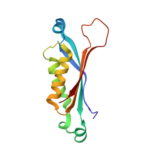
    Potent inhibitors of 7,8-dihydroneopterin aldolase (DHNA; EC 4.1.2.25) have been discovered using CrystaLEAD X-ray crystallographic high-throughput screening followed by structure-directed optimization. Screening of a 10 000 compound random library provided several low affinity leads and their corresponding X-ray crystal structures bound to the enzyme. The presence of a common structural feature in each of the leads suggested a strategy for the construction of a directed library of approximately 1000 compounds that were screened for inhibitory activity in a traditional enzyme assay. Several lead compounds with IC(50) values of about 1 microM against DHNA were identified, and crystal structures of their enzyme-bound complexes were obtained by cocrystallization. Structure-directed optimization of one of the leads thus identified afforded potent inhibitors with submicromolar IC(50) values.


  • Organizational Affiliation
    • Infectious Disease Research, Abbott Laboratories, 200 Abbott Park Road, Abbott Park, Illinois 60064-6217, USA. will.sanders@abbott.com

Macromolecule Content 

  • Total Structure Weight: 14.02 kDa 
  • Atom Count: 985 
  • Modeled Residue Count: 121 
  • Deposited Residue Count: 121 
  • Unique protein chains: 1

Macromolecules

Find similar proteins by:|  3D Structure
Entity ID: 1
MoleculeChains  Sequence LengthOrganismDetailsImage
Dihydroneopterin aldolase121Staphylococcus aureusMutation(s): 0 
Gene Names: folB
EC: 4.1.2.25 (PDB Primary Data), 5.1.99.8 (UniProt)
UniProt
Find proteins for P56740 (Staphylococcus aureus)
Explore P56740 
Go to UniProtKB:  P56740
Entity Groups
Sequence Clusters30% Identity50% Identity70% Identity90% Identity95% Identity100% Identity
UniProt GroupP56740
Sequence Annotations
Expand
Reference Sequence

Small Molecules

Ligands 1 Unique
IDChains Name / Formula / InChI Key2D Diagram3D Interactions
NPR

Query on NPR


Download:Ideal Coordinates CCD File
B [auth A]2-AMINO-7,8-DIHYDRO-6-(1,2,3-TRIHYDROXYPROPYL)-4(1H)-PTERIDINONE
C9 H13 N5 O4
YQIFAMYNGGOTFB-NJGYIYPDSA-N

Experimental Data & Validation

Experimental Data

  • Method: X-RAY DIFFRACTION
  • Resolution: 2.40 Å
  • R-Value Free:  0.368 (Depositor) 
  • R-Value Work:  0.279 (Depositor) 
Space Group: I 4 2 2
Unit Cell:
Length ( Å )Angle ( ˚ )
a = 60.947α = 90
b = 60.947β = 90
c = 123.689γ = 90
Software Package:
Software NamePurpose
DENZOdata reduction
SCALEPACKdata scaling
CNSrefinement
CNSphasing

Structure Validation

View Full Validation Report


Entry History 

Revision History  (Full details and data files)

  • Version 1.0: 2004-10-19
    Type: Initial release
  • Version 1.1: 2008-04-30
    Changes: Version format compliance
  • Version 1.2: 2011-07-13
    Changes: Derived calculations, Version format compliance
  • Version 1.3: 2024-12-25
    Changes: Advisory, Data collection, Database references, Derived calculations, Structure summary