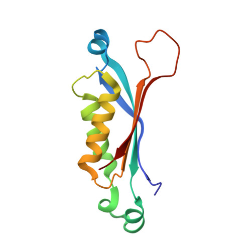
1RSI

DHNA complex with 2-Amino-5-bromo-3-hydroxy-6-phenylpyrimidine


Experimental Data Snapshot

  • Method: X-RAY DIFFRACTION
  • Resolution: 2.20 Å
  • R-Value Free: 0.339 
  • R-Value Work: 0.276 

wwPDB Validation   3D Report Full Report


This is version 1.3 of the entry. See complete history


Literature

Discovery of Potent Inhibitors of Dihydroneopterin Aldolase Using CrystaLEAD High-Throughput X-ray Crystallographic Screening and Structure-Directed Lead Optimization.

Sanders, W.J.Nienaber, V.L.Lerner, C.G.McCall, J.O.Merrick, S.M.Swanson, S.J.Harlan, J.E.Stoll, V.S.Stamper, G.F.Betz, S.F.Condroski, K.R.Meadows, R.P.Severin, J.M.Walter, K.A.Magdalinos, P.Jakob, C.G.Wagner, R.Beutel, B.A.

(2004) J Med Chem 47: 1709-1718

  • DOI: https://doi.org/10.1021/jm030497y
  • Primary Citation of Related Structures:  
    1RRI, 1RRW, 1RRY, 1RS2, 1RS4, 1RSD, 1RSI, 1U68

  • PubMed Abstract: 

    Potent inhibitors of 7,8-dihydroneopterin aldolase (DHNA; EC 4.1.2.25) have been discovered using CrystaLEAD X-ray crystallographic high-throughput screening followed by structure-directed optimization. Screening of a 10 000 compound random library provided several low affinity leads and their corresponding X-ray crystal structures bound to the enzyme. The presence of a common structural feature in each of the leads suggested a strategy for the construction of a directed library of approximately 1000 compounds that were screened for inhibitory activity in a traditional enzyme assay. Several lead compounds with IC(50) values of about 1 microM against DHNA were identified, and crystal structures of their enzyme-bound complexes were obtained by cocrystallization. Structure-directed optimization of one of the leads thus identified afforded potent inhibitors with submicromolar IC(50) values.


  • Organizational Affiliation

    Infectious Disease Research, Abbott Laboratories, 200 Abbott Park Road, Abbott Park, Illinois 60064-6217, USA. will.sanders@abbott.com


Macromolecules
Find similar proteins by:  (by identity cutoff)  |  3D Structure
Entity ID: 1
MoleculeChains Sequence LengthOrganismDetailsImage
Dihydroneopterin aldolase121Staphylococcus aureusMutation(s): 0 
Gene Names: FOLB
EC: 4.1.2.25 (PDB Primary Data), 5.1.99.8 (UniProt)
UniProt
Find proteins for P56740 (Staphylococcus aureus)
Explore P56740 
Go to UniProtKB:  P56740
Entity Groups  
Sequence Clusters30% Identity50% Identity70% Identity90% Identity95% Identity100% Identity
UniProt GroupP56740
Sequence Annotations
Expand
  • Reference Sequence
Small Molecules
Ligands 1 Unique
IDChains Name / Formula / InChI Key2D Diagram3D Interactions
977
Query on 977

Download Ideal Coordinates CCD File 
B [auth A]2-AMINO-5-BROMO-6-PHENYLPYRIMIDIN-4-OL
C10 H8 Br N3 O
CIUUIPMOFZIWIZ-UHFFFAOYSA-N
Binding Affinity Annotations 
IDSourceBinding Affinity
977 PDBBind:  1RSI IC50: 3.80e+4 (nM) from 1 assay(s)
Experimental Data & Validation

Experimental Data

  • Method: X-RAY DIFFRACTION
  • Resolution: 2.20 Å
  • R-Value Free: 0.339 
  • R-Value Work: 0.276 
  • Space Group: I 4 2 2
Unit Cell:
Length ( Å )Angle ( ˚ )
a = 60.947α = 90
b = 60.947β = 90
c = 123.689γ = 90
Software Package:
Software NamePurpose
CNXrefinement
HKL-2000data reduction
SCALEPACKdata scaling
X-PLORphasing

Structure Validation

View Full Validation Report


Entry History 

Revision History  (Full details and data files)

  • Version 1.0: 2004-03-30
    Type: Initial release
  • Version 1.1: 2008-04-29
    Changes: Version format compliance
  • Version 1.2: 2011-07-13
    Changes: Derived calculations, Version format compliance
  • Version 1.3: 2024-02-14
    Changes: Data collection, Database references, Derived calculations