1U2O

Crystal Structure Of The N-Domain Of Grp94 Lacking The Charged Domain In Complex With Neca


Experimental Data Snapshot

  • Method: X-RAY DIFFRACTION
  • Resolution: 2.10 Å
  • R-Value Free: 0.257 
  • R-Value Work: 0.213 
  • R-Value Observed: 0.213 

wwPDB Validation   3D Report Full Report


Ligand Structure Quality Assessment 


This is version 1.4 of the entry. See complete history


Literature

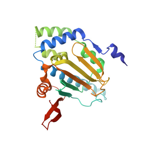
Structure of the N-terminal domain of GRP94. Basis for ligand specificity and regulation

Soldano, K.L.Jivan, A.Nicchitta, C.V.Gewirth, D.T.

(2003) J Biol Chem 278: 48330-48338

  • DOI: https://doi.org/10.1074/jbc.M308661200
  • Primary Citation of Related Structures:  
    1QY8, 1QYE, 1U2O, 6D28

  • PubMed Abstract: 

    GRP94, the endoplasmic reticulum (ER) paralog of the chaperone Hsp90, plays an essential role in the structural maturation or secretion of a subset of proteins destined for transport to the cell surface, such as the Toll-like receptors 2 and 4, and IgG, respectively. GRP94 differs from cytoplasmic Hsp90 by exhibiting very weak ATP binding and hydrolysis activity. GRP94 also binds selectively to a series of substituted adenosine analogs. The high resolution crystal structures at 1.75-2.1 A of the N-terminal and adjacent charged domains of GRP94 in complex with N-ethylcarboxamidoadenosine, radicicol, and 2-chlorodideoxyadenosine reveals a structural mechanism for ligand discrimination among hsp90 family members. The structures also identify a putative subdomain that may act as a ligand-responsive switch. The residues of the charged region fold into a disordered loop whose termini are ordered and continue the twisted beta sheet that forms the structural core of the N-domain. This continuation of the beta sheet past the charged domain suggests a structural basis for the association of the N-terminal and middle domains of the full-length chaperone.


  • Organizational Affiliation

    Departments of Biochemistry and Cell Biology, Duke University Medical Center, Durham, North Carolina 27710, USA.


Macromolecules
Find similar proteins by:  (by identity cutoff)  |  3D Structure
Entity ID: 1
MoleculeChains Sequence LengthOrganismDetailsImage
Endoplasmin
A, B
236Canis lupus familiarisMutation(s): 0 
Gene Names: TRA1
UniProt
Find proteins for P41148 (Canis lupus familiaris)
Explore P41148 
Go to UniProtKB:  P41148
Entity Groups  
Sequence Clusters30% Identity50% Identity70% Identity90% Identity95% Identity100% Identity
UniProt GroupP41148
Sequence Annotations
Expand
  • Reference Sequence
Small Molecules
Ligands 3 Unique
IDChains Name / Formula / InChI Key2D Diagram3D Interactions
NEC
Query on NEC

Download Ideal Coordinates CCD File 
C [auth A],
F [auth B]
N-ETHYL-5'-CARBOXAMIDO ADENOSINE
C12 H16 N6 O4
JADDQZYHOWSFJD-FLNNQWSLSA-N
1PE
Query on 1PE

Download Ideal Coordinates CCD File 
G [auth B]PENTAETHYLENE GLYCOL
C10 H22 O6
JLFNLZLINWHATN-UHFFFAOYSA-N
PG4
Query on PG4

Download Ideal Coordinates CCD File 
D [auth A],
E [auth A],
H [auth B],
I [auth B],
J [auth B]
TETRAETHYLENE GLYCOL
C8 H18 O5
UWHCKJMYHZGTIT-UHFFFAOYSA-N
Experimental Data & Validation

Experimental Data

  • Method: X-RAY DIFFRACTION
  • Resolution: 2.10 Å
  • R-Value Free: 0.257 
  • R-Value Work: 0.213 
  • R-Value Observed: 0.213 
  • Space Group: P 21 21 21
Unit Cell:
Length ( Å )Angle ( ˚ )
a = 65.3α = 90
b = 84.2β = 90
c = 94.091γ = 90
Software Package:
Software NamePurpose
CNSrefinement
HKL-2000data reduction
SCALEPACKdata scaling
CNSphasing

Structure Validation

View Full Validation Report


Ligand Structure Quality Assessment 


Entry History 

Deposition Data

Revision History  (Full details and data files)

  • Version 1.0: 2004-08-03
    Type: Initial release
  • Version 1.1: 2008-04-30
    Changes: Version format compliance
  • Version 1.2: 2011-07-13
    Changes: Version format compliance
  • Version 1.3: 2017-08-23
    Changes: Source and taxonomy
  • Version 1.4: 2023-10-25
    Changes: Data collection, Database references, Derived calculations, Refinement description