1TW1

beta-1,4-galactosyltransferase mutant Met344His (m344H-Gal-T1) complex with UDP-galactose and magnesium


Experimental Data Snapshot

  • Method: X-RAY DIFFRACTION
  • Resolution: 2.30 Å
  • R-Value Free: 0.261 
  • R-Value Work: 0.196 
  • R-Value Observed: 0.196 

wwPDB Validation   3D Report Full Report


Ligand Structure Quality Assessment 


This is version 1.4 of the entry. See complete history


Literature

Effect of the Met344His mutation on the conformational dynamics of bovine beta-1,4-galactosyltransferase: crystal structure of the Met344His mutant in complex with chitobiose

Ramakrishnan, B.Boeggeman, E.Qasba, P.K.

(2004) Biochemistry 43: 12513-12522

  • DOI: https://doi.org/10.1021/bi049007+
  • Primary Citation of Related Structures:  
    1TVY, 1TW1, 1TW5

  • PubMed Abstract: 

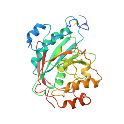
    Beta-1,4-galactosyltransferase (beta4Gal-T1) in the presence of manganese ion transfers galactose from UDP-galactose (UDP-Gal) to N-acetylglucosamine (GlcNAc) that is either free or linked to an oligosaccharide. Crystallographic studies on bovine beta4Gal-T1 have shown that the primary metal binding site is located in the hinge region of a long flexible loop, which upon Mn(2+) and UDP-Gal binding changes from an open to a closed conformation. This conformational change creates an oligosaccharide binding site in the enzyme. Neither UDP nor UDP analogues efficiently induce these conformational changes in the wild-type enzyme, thereby restricting the structural analysis of the acceptor binding site. The binding of Mn(2+) involves an uncommon coordination to the Sdelta atom of Met344; when it is mutated to His, the mutant M344H, in the presence of Mn(2+) and UDP-hexanolamine, readily changes to a closed conformation, facilitating the structural analysis of the enzyme bound with an oligosaccharide acceptor. Although the mutant M344H loses 98% of its Mn(2+)-dependent activity, it exhibits 25% of its activity in the presence of Mg(2+). The crystal structures of M344H-Gal-T1 in complex with either UDP-Gal.Mn(2+) or UDP-Gal.Mg(2+), determined at 2.3 A resolution, show that the mutant enzyme in these complexes is in a closed conformation, and the coordination stereochemistry of Mg(2+) is quite similar to that of Mn(2+). Although either Mn(2+) or Mg(2+), together with UDP-Gal, binds and changes the conformation of the M344H mutant to the closed one, it is the Mg(2+) complex that engages efficiently in catalyses. Thus, this property enabled us to crystallize the M344H mutant for the first time with the acceptor substrate chitobiose in the presence of UDP-hexanolamine and Mn(2+). The crystal structure determined at 2.3 A resolution reveals that the GlcNAc residue at the nonreducing end of chitobiose makes extensive hydrophobic interactions with the highly conserved Tyr286 residue.


  • Organizational Affiliation

    Structural Glycobiology Section, Laboratory of Experimental and Computational Biology, SAIC-Frederick, Inc., Center for Cancer Research, National Cancer Institute, Frederick, Maryland 21702-1201, USA.


Macromolecules
Find similar proteins by:  (by identity cutoff)  |  3D Structure
Entity ID: 1
MoleculeChains Sequence LengthOrganismDetailsImage
Beta-1,4-galactosyltransferase 1
A, B
286Bos taurusMutation(s): 2 
Gene Names: B4GALT1
EC: 2.4.1.22 (PDB Primary Data), 2.4.1.90 (PDB Primary Data), 2.4.1.38 (PDB Primary Data)
UniProt
Find proteins for P08037 (Bos taurus)
Explore P08037 
Go to UniProtKB:  P08037
Entity Groups  
Sequence Clusters30% Identity50% Identity70% Identity90% Identity95% Identity100% Identity
UniProt GroupP08037
Sequence Annotations
Expand
  • Reference Sequence
Small Molecules
Ligands 5 Unique
IDChains Name / Formula / InChI Key2D Diagram3D Interactions
GDU
Query on GDU

Download Ideal Coordinates CCD File 
C [auth A],
I [auth B]
GALACTOSE-URIDINE-5'-DIPHOSPHATE
C15 H24 N2 O17 P2
HSCJRCZFDFQWRP-ABVWGUQPSA-N
MES
Query on MES

Download Ideal Coordinates CCD File 
N [auth B]2-(N-MORPHOLINO)-ETHANESULFONIC ACID
C6 H13 N O4 S
SXGZJKUKBWWHRA-UHFFFAOYSA-N
SO4
Query on SO4

Download Ideal Coordinates CCD File 
E [auth A],
F [auth A],
K [auth B],
L [auth B],
M [auth B]
SULFATE ION
O4 S
QAOWNCQODCNURD-UHFFFAOYSA-L
DIO
Query on DIO

Download Ideal Coordinates CCD File 
G [auth A],
H [auth A]
1,4-DIETHYLENE DIOXIDE
C4 H8 O2
RYHBNJHYFVUHQT-UHFFFAOYSA-N
MG
Query on MG

Download Ideal Coordinates CCD File 
D [auth A],
J [auth B]
MAGNESIUM ION
Mg
JLVVSXFLKOJNIY-UHFFFAOYSA-N
Experimental Data & Validation

Experimental Data

  • Method: X-RAY DIFFRACTION
  • Resolution: 2.30 Å
  • R-Value Free: 0.261 
  • R-Value Work: 0.196 
  • R-Value Observed: 0.196 
  • Space Group: P 21 21 21
Unit Cell:
Length ( Å )Angle ( ˚ )
a = 50.006α = 90
b = 91.517β = 90
c = 142.751γ = 90
Software Package:
Software NamePurpose
HKL-2000data collection
SCALEPACKdata scaling
AMoREphasing
CNSrefinement
HKL-2000data reduction

Structure Validation

View Full Validation Report


Ligand Structure Quality Assessment 


Entry History 

Deposition Data

Revision History  (Full details and data files)

  • Version 1.0: 2004-12-14
    Type: Initial release
  • Version 1.1: 2008-04-30
    Changes: Version format compliance
  • Version 1.2: 2011-07-13
    Changes: Version format compliance
  • Version 1.3: 2021-10-27
    Changes: Database references, Derived calculations
  • Version 1.4: 2023-08-23
    Changes: Data collection, Refinement description