1TA0 | pdb_00001ta0

Three-dimensional structure of a RNA-polymerase II binding protein with associated ligand.


Experimental Data Snapshot

  • Method: X-RAY DIFFRACTION
  • Resolution: 2.10 Å
  • R-Value Free: 
    0.227 (Depositor), 0.221 (DCC) 
  • R-Value Work: 
    0.207 (Depositor), 0.199 (DCC) 

wwPDB Validation 3D Report Full Report

Validation slider image for 1TA0

This is version 1.3 of the entry. See complete history

Literature

Structure and Mechanism of RNA Polymerase II CTD Phosphatases.

Kamenski, T.Heilmeier, S.Meinhart, T.Cramer, P.

(2004) Mol Cell 15: 399-407

  • DOI: https://doi.org/10.1016/j.molcel.2004.06.035
  • Primary Citation Related Structures: 
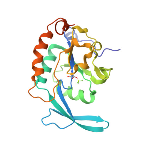
    1T9Z, 1TA0

  • PubMed Abstract: 

    Recycling of RNA polymerase II (Pol II) after transcription requires dephosphorylation of the polymerase C-terminal domain (CTD) by the phosphatase Fcp1. We report the X-ray structure of the small CTD phosphatase Scp1, which is homologous to the Fcp1 catalytic domain. The structure shows a core fold and an active center similar to those of phosphotransferases and phosphohydrolases that solely share a DXDX(V/T) signature motif with Fcp1/Scp1. We demonstrate that the first aspartate in the signature motif undergoes metal-assisted phosphorylation during catalysis, resulting in a phosphoaspartate intermediate that was structurally mimicked with the inhibitor beryllofluoride. Specificity may result from CTD binding to a conserved hydrophobic pocket between the active site and an insertion domain that is unique to Fcp1/Scp1. Fcp1 specificity may additionally arise from phosphatase recruitment near the CTD via the Pol II subcomplex Rpb4/7, which is shown to be required for binding of Fcp1 to the polymerase in vitro.


  • Organizational Affiliation
    • Department of Chemistry and Biochemistry, Gene Center, University of Munich, Feodor-Lynen-Str. 25, 81377 Munich, Germany.

Macromolecule Content 

  • Total Structure Weight: 22.93 kDa 
  • Atom Count: 1,637 
  • Modeled Residue Count: 181 
  • Deposited Residue Count: 197 
  • Unique protein chains: 1

Macromolecules

Find similar proteins by:|  3D Structure
Entity ID: 1
MoleculeChains  Sequence LengthOrganismDetailsImage
Carboxy-terminal domain RNA polymerase II polypeptide A small phosphatase 1197Homo sapiensMutation(s): 1 
Gene Names: CTDSP1NIF3NLIIF
EC: 3.1.3.16
UniProt & NIH Common Fund Data Resources
Find proteins for Q9GZU7 (Homo sapiens)
Explore Q9GZU7 
Go to UniProtKB:  Q9GZU7
PHAROS:  Q9GZU7
GTEx:  ENSG00000144579 
Entity Groups
Sequence Clusters30% Identity50% Identity70% Identity90% Identity95% Identity100% Identity
UniProt GroupQ9GZU7
Sequence Annotations
Expand
Reference Sequence

Small Molecules

Modified Residues  1 Unique
IDChains TypeFormula2D DiagramParent
BFD
Query on BFD
A
L-PEPTIDE LINKINGC4 H6 Be F3 N O4ASP

Experimental Data & Validation

Experimental Data

  • Method: X-RAY DIFFRACTION
  • Resolution: 2.10 Å
  • R-Value Free:  0.227 (Depositor), 0.221 (DCC) 
  • R-Value Work:  0.207 (Depositor), 0.199 (DCC) 
Space Group: P 21 21 2
Unit Cell:
Length ( Å )Angle ( ˚ )
a = 117.82α = 90
b = 47.17β = 90
c = 40.07γ = 90
Software Package:
Software NamePurpose
XDSdata scaling
XDSdata reduction
CNSrefinement
CNSphasing

Structure Validation

View Full Validation Report


Entry History 

Deposition Data

Revision History  (Full details and data files)

  • Version 1.0: 2004-08-31
    Type: Initial release
  • Version 1.1: 2008-04-30
    Changes: Version format compliance
  • Version 1.2: 2011-07-13
    Changes: Version format compliance
  • Version 1.3: 2024-10-16
    Changes: Data collection, Database references, Derived calculations, Structure summary