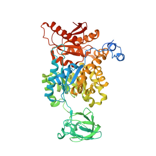
1T5A

Human Pyruvate Kinase M2


Experimental Data Snapshot

  • Method: X-RAY DIFFRACTION
  • Resolution: 2.80 Å
  • R-Value Free: 0.278 
  • R-Value Work: 0.231 

Starting Model: experimental
View more details

wwPDB Validation   3D Report Full Report


Ligand Structure Quality Assessment 


This is version 1.5 of the entry. See complete history


Literature

Structural basis for tumor pyruvate kinase M2 allosteric regulation and catalysis.

Dombrauckas, J.D.Santarsiero, B.D.Mesecar, A.D.

(2005) Biochemistry 44: 9417-9429

  • DOI: https://doi.org/10.1021/bi0474923
  • Primary Citation of Related Structures:  
    1T5A

  • PubMed Abstract: 

    Four isozymes of pyruvate kinase are differentially expressed in human tissue. Human pyruvate kinase isozyme M2 (hPKM2) is expressed in early fetal tissues and is progressively replaced by the other three isozymes, M1, R, and L, immediately after birth. In most cancer cells, hPKM2 is once again expressed to promote tumor cell proliferation. Because of its almost ubiquitous presence in cancer cells, hPKM2 has been designated as tumor specific PK-M2, and its presence in human plasma is currently being used as a molecular marker for the diagnosis of various cancers. The X-ray structure of human hPKM2 complexed with Mg(2+), K(+), the inhibitor oxalate, and the allosteric activator fructose 1,6-bisphosphate (FBP) has been determined to a resolution of 2.82 A. The active site of hPKM2 is in a partially closed conformation most likely resulting from a ligand-induced domain closure promoted by the binding of FBP. In all four subunits of the enzyme tetramer, a conserved water molecule is observed on the 2-si face of the prospective enolate and supports the hypothesis that a proton-relay system is acting as the proton donor of the reaction (1). Significant structural differences among the human M2, rabbit muscle M1, and the human R isozymes are observed, especially in the orientation of the FBP-activating loop, which is in a closed conformation when FBP is bound. The structural differences observed between the PK isozymes could potentially be exploited as unique structural templates for the design of allosteric drugs against the disease states associated with the various PK isozymes, especially cancer and nonspherocytic hemolytic anemia.


  • Organizational Affiliation

    Center for Pharmaceutical Biotechnology and Department of Medicinal Chemistry and Pharmacognosy, University of Illinois at Chicago, Chicago, Illinois 60607, USA.


Macromolecules
Find similar proteins by:  (by identity cutoff)  |  3D Structure
Entity ID: 1
MoleculeChains Sequence LengthOrganismDetailsImage
Pyruvate kinase, M2 isozyme
A, B, C, D
567Homo sapiensMutation(s): 0 
Gene Names: PKM2PKM
EC: 2.7.1.40
UniProt & NIH Common Fund Data Resources
Find proteins for P14618 (Homo sapiens)
Explore P14618 
Go to UniProtKB:  P14618
PHAROS:  P14618
GTEx:  ENSG00000067225 
Entity Groups  
Sequence Clusters30% Identity50% Identity70% Identity90% Identity95% Identity100% Identity
UniProt GroupP14618
Sequence Annotations
Expand
  • Reference Sequence
Small Molecules
Ligands 6 Unique
IDChains Name / Formula / InChI Key2D Diagram3D Interactions
FBP
Query on FBP

Download Ideal Coordinates CCD File 
E [auth A],
IA [auth D],
O [auth B],
X [auth C]
1,6-di-O-phosphono-beta-D-fructofuranose
C6 H14 O12 P2
RNBGYGVWRKECFJ-ARQDHWQXSA-N
PO4
Query on PO4

Download Ideal Coordinates CCD File 
G [auth A],
KA [auth D],
Q [auth B],
Z [auth C]
PHOSPHATE ION
O4 P
NBIIXXVUZAFLBC-UHFFFAOYSA-K
GOL
Query on GOL

Download Ideal Coordinates CCD File 
CA [auth C]
DA [auth C]
EA [auth C]
FA [auth C]
GA [auth C]
CA [auth C],
DA [auth C],
EA [auth C],
FA [auth C],
GA [auth C],
HA [auth C],
J [auth A],
K [auth A],
L [auth A],
M [auth A],
N [auth A],
NA [auth D],
OA [auth D],
PA [auth D],
QA [auth D],
RA [auth D],
T [auth B],
U [auth B],
V [auth B],
W [auth B]
GLYCEROL
C3 H8 O3
PEDCQBHIVMGVHV-UHFFFAOYSA-N
OXL
Query on OXL

Download Ideal Coordinates CCD File 
F [auth A],
JA [auth D],
P [auth B],
Y [auth C]
OXALATE ION
C2 O4
MUBZPKHOEPUJKR-UHFFFAOYSA-L
K
Query on K

Download Ideal Coordinates CCD File 
BA [auth C],
I [auth A],
MA [auth D],
S [auth B]
POTASSIUM ION
K
NPYPAHLBTDXSSS-UHFFFAOYSA-N
MG
Query on MG

Download Ideal Coordinates CCD File 
AA [auth C],
H [auth A],
LA [auth D],
R [auth B]
MAGNESIUM ION
Mg
JLVVSXFLKOJNIY-UHFFFAOYSA-N
Binding Affinity Annotations 
IDSourceBinding Affinity
OXL PDBBind:  1T5A Ki: 2.40e+4 (nM) from 1 assay(s)
Experimental Data & Validation

Experimental Data

  • Method: X-RAY DIFFRACTION
  • Resolution: 2.80 Å
  • R-Value Free: 0.278 
  • R-Value Work: 0.231 
  • Space Group: P 21 21 21
Unit Cell:
Length ( Å )Angle ( ˚ )
a = 108.209α = 90
b = 145.006β = 90
c = 159.26γ = 90
Software Package:
Software NamePurpose
DENZOdata reduction
SCALEPACKdata scaling
EPMRphasing
CNSrefinement

Structure Validation

View Full Validation Report


Ligand Structure Quality Assessment 


Entry History 

Deposition Data

Revision History  (Full details and data files)

  • Version 1.0: 2005-07-12
    Type: Initial release
  • Version 1.1: 2008-04-30
    Changes: Version format compliance
  • Version 1.2: 2011-07-13
    Changes: Non-polymer description, Version format compliance
  • Version 1.3: 2020-07-29
    Type: Remediation
    Reason: Carbohydrate remediation
    Changes: Data collection, Database references, Derived calculations, Structure summary
  • Version 1.4: 2022-12-21
    Changes: Database references, Structure summary
  • Version 1.5: 2023-09-20
    Changes: Data collection, Refinement description