1SYQ

Human vinculin head domain VH1, residues 1-258, in complex with human talin's vinculin binding site 1, residues 607-636


Experimental Data Snapshot

  • Method: X-RAY DIFFRACTION
  • Resolution: 2.42 Å
  • R-Value Free: 0.236 
  • R-Value Work: 0.188 
  • R-Value Observed: 0.194 

wwPDB Validation   3D Report Full Report


This is version 1.3 of the entry. See complete history


Literature

Structural basis for amplifying vinculin activation by talin

Izard, T.Vonrhein, C.

(2004) J Biol Chem 279: 27667-27678

  • DOI: https://doi.org/10.1074/jbc.M403076200
  • Primary Citation of Related Structures:  
    1SYQ

  • PubMed Abstract: 

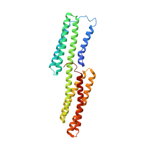
    Talin interactions with vinculin are essential for focal adhesions. Curiously, talin contains three noncontiguous vinculin binding sites (VBS) that can bind individually to the vinculin head (Vh) domain. Here we report the crystal structure of the human Vh.VBS1 complex, a validated model of the Vh.VBS2 structure, and biochemical studies that demonstrate that all of talin VBSs activate vinculin by provoking helical bundle conversion of the Vh domain, which displaces the vinculin tail (Vt) domain. Thus, helical bundle conversion is a structurally conserved response in talin-vinculin interactions. Furthermore, talin VBSs bind to Vh in a mutually exclusive manner but do differ in their affinity for Vh and in their ability to displace Vt, suggesting that the strengths of these interactions could lead to differences in signaling outcome. These findings support a model in which talin binds to and activates multiple vinculin molecules to provoke rapid reorganization of the actin cytoskeleton.


  • Organizational Affiliation

    Department of Hematology-Oncology, St. Jude Children's Research Hospital, Memphis, Tennessee 38105, USA. tina.izard@stjude.org


Macromolecules
Find similar proteins by:  (by identity cutoff)  |  3D Structure
Entity ID: 1
MoleculeChains Sequence LengthOrganismDetailsImage
vinculin isoform VCL264Homo sapiensMutation(s): 0 
UniProt & NIH Common Fund Data Resources
Find proteins for P18206 (Homo sapiens)
Explore P18206 
Go to UniProtKB:  P18206
PHAROS:  P18206
GTEx:  ENSG00000035403 
Entity Groups  
Sequence Clusters30% Identity50% Identity70% Identity90% Identity95% Identity100% Identity
UniProt GroupP18206
Sequence Annotations
Expand
  • Reference Sequence

Find similar proteins by:  Sequence   |   3D Structure  

Entity ID: 2
MoleculeChains Sequence LengthOrganismDetailsImage
Talin 125Homo sapiensMutation(s): 0 
Gene Names: TLN1
UniProt & NIH Common Fund Data Resources
Find proteins for Q9Y490 (Homo sapiens)
Explore Q9Y490 
Go to UniProtKB:  Q9Y490
PHAROS:  Q9Y490
GTEx:  ENSG00000137076 
Entity Groups  
Sequence Clusters30% Identity50% Identity70% Identity90% Identity95% Identity100% Identity
UniProt GroupQ9Y490
Sequence Annotations
Expand
  • Reference Sequence
Experimental Data & Validation

Experimental Data

  • Method: X-RAY DIFFRACTION
  • Resolution: 2.42 Å
  • R-Value Free: 0.236 
  • R-Value Work: 0.188 
  • R-Value Observed: 0.194 
  • Space Group: H 3 2
Unit Cell:
Length ( Å )Angle ( ˚ )
a = 141.098α = 90
b = 141.098β = 90
c = 104.92γ = 120
Software Package:
Software NamePurpose
CNSrefinement
HKL-2000data reduction
HKL-2000data scaling

Structure Validation

View Full Validation Report


Entry History 

Deposition Data

Revision History  (Full details and data files)

  • Version 1.0: 2004-07-20
    Type: Initial release
  • Version 1.1: 2008-04-30
    Changes: Version format compliance
  • Version 1.2: 2011-07-13
    Changes: Version format compliance
  • Version 1.3: 2024-02-14
    Changes: Data collection, Database references