1SXJ | pdb_00001sxj

Crystal Structure of the Eukaryotic Clamp Loader (Replication Factor C, RFC) Bound to the DNA Sliding Clamp (Proliferating Cell Nuclear Antigen, PCNA)


Experimental Data Snapshot

  • Method: X-RAY DIFFRACTION
  • Resolution: 2.85 Å
  • R-Value Free: 
    0.306 (Depositor), 0.300 (DCC) 
  • R-Value Work: 
    0.251 (Depositor), 0.240 (DCC) 
  • R-Value Observed: 
    0.251 (Depositor) 

Starting Models: experimental
View more details

wwPDB Validation   3D Report Full Report


Ligand Structure Quality Assessment 

Created with Raphaël 2.3.0Worse 01 BetterLigand structure goodness of fit to experimental dataBest fitted AGSClick on this verticalbar to view detailsBest fitted ADPClick on this verticalbar to view details

This is version 1.6 of the entry. See complete history


Literature

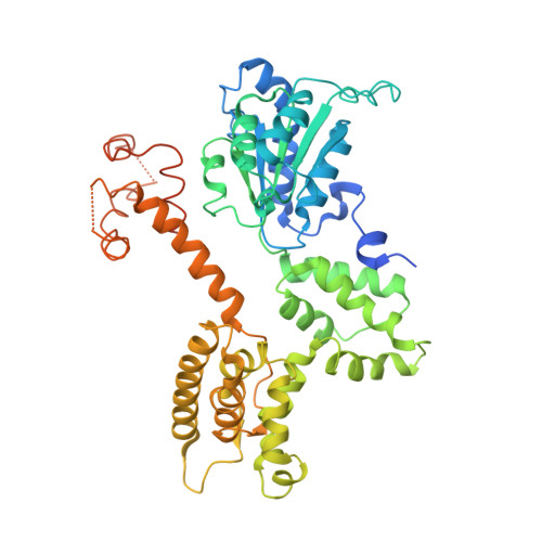
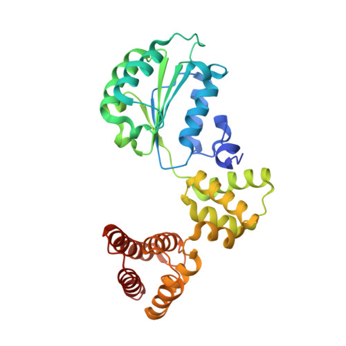
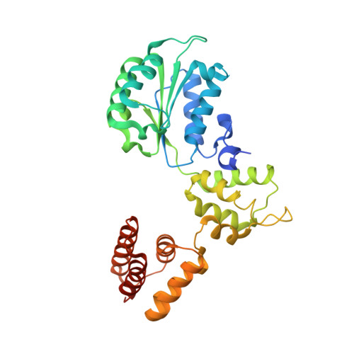
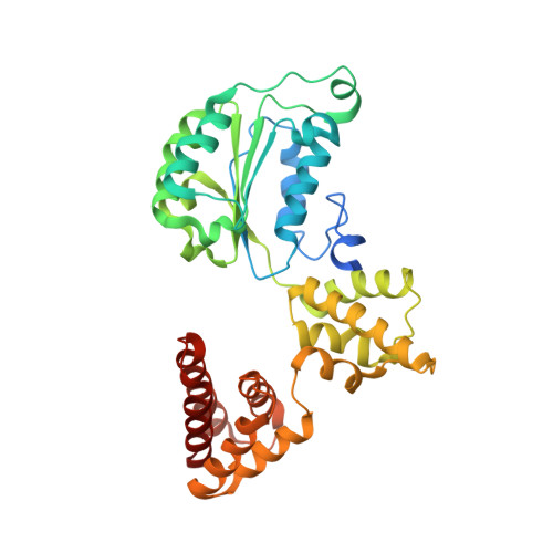
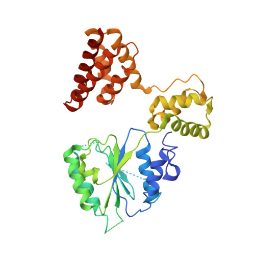
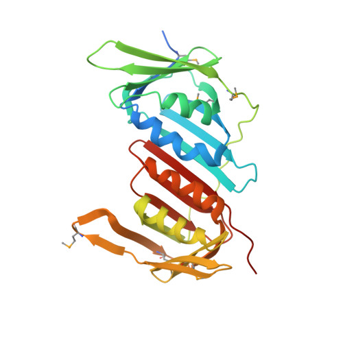
Structural analysis of a eukaryotic sliding DNA clamp-clamp loader complex.

Bowman, G.D.O'Donnell, M.Kuriyan, J.

(2004) Nature 429: 724-730

  • DOI: https://doi.org/10.1038/nature02585
  • Primary Citation of Related Structures:  
    1SXJ

  • PubMed Abstract: 

    Sliding clamps are ring-shaped proteins that encircle DNA and confer high processivity on DNA polymerases. Here we report the crystal structure of the five-protein clamp loader complex (replication factor-C, RFC) of the yeast Saccharomyces cerevisiae, bound to the sliding clamp (proliferating cell nuclear antigen, PCNA). Tight interfacial coordination of the ATP analogue ATP-gammaS by RFC results in a spiral arrangement of the ATPase domains of the clamp loader above the PCNA ring. Placement of a model for primed DNA within the central hole of PCNA reveals a striking correspondence between the RFC spiral and the grooves of the DNA double helix. This model, in which the clamp loader complex locks onto primed DNA in a screw-cap-like arrangement, provides a simple explanation for the process by which the engagement of primer-template junctions by the RFC:PCNA complex results in ATP hydrolysis and release of the sliding clamp on DNA.


  • Organizational Affiliation

    Howard Hughes Medical Institute, Department of Molecular and Cell Biology and Department of Chemistry, University of California, Berkeley, California 94720, USA.


Macromolecules
Find similar proteins by:  (by identity cutoff)  |  3D Structure
Entity ID: 1
MoleculeChains Sequence LengthOrganismDetailsImage
Activator 1 95 kDa subunit516Saccharomyces cerevisiaeMutation(s): 0 
Gene Names: RFC1CDC44YOR217WYOR50-7
UniProt
Find proteins for P38630 (Saccharomyces cerevisiae (strain ATCC 204508 / S288c))
Explore P38630 
Go to UniProtKB:  P38630
Entity Groups  
Sequence Clusters30% Identity50% Identity70% Identity90% Identity95% Identity100% Identity
UniProt GroupP38630
Sequence Annotations
Expand
  • Reference Sequence
Find similar proteins by:  (by identity cutoff)  |  3D Structure
Entity ID: 2
MoleculeChains Sequence LengthOrganismDetailsImage
Activator 1 37 kDa subunit323Saccharomyces cerevisiaeMutation(s): 1 
Gene Names: RFC4YOL094CO0923
UniProt
Find proteins for P40339 (Saccharomyces cerevisiae (strain ATCC 204508 / S288c))
Explore P40339 
Go to UniProtKB:  P40339
Entity Groups  
Sequence Clusters30% Identity50% Identity70% Identity90% Identity95% Identity100% Identity
UniProt GroupP40339
Sequence Annotations
Expand
  • Reference Sequence
Find similar proteins by:  (by identity cutoff)  |  3D Structure
Entity ID: 3
MoleculeChains Sequence LengthOrganismDetailsImage
Activator 1 40 kDa subunit340Saccharomyces cerevisiaeMutation(s): 1 
Gene Names: RFC3YNL290WN0533
UniProt
Find proteins for P38629 (Saccharomyces cerevisiae (strain ATCC 204508 / S288c))
Explore P38629 
Go to UniProtKB:  P38629
Entity Groups  
Sequence Clusters30% Identity50% Identity70% Identity90% Identity95% Identity100% Identity
UniProt GroupP38629
Sequence Annotations
Expand
  • Reference Sequence
Find similar proteins by:  (by identity cutoff)  |  3D Structure
Entity ID: 4
MoleculeChains Sequence LengthOrganismDetailsImage
Activator 1 41 kDa subunit353Saccharomyces cerevisiaeMutation(s): 1 
Gene Names: RFC2YJR068WJ1808
UniProt
Find proteins for P40348 (Saccharomyces cerevisiae (strain ATCC 204508 / S288c))
Explore P40348 
Go to UniProtKB:  P40348
Entity Groups  
Sequence Clusters30% Identity50% Identity70% Identity90% Identity95% Identity100% Identity
UniProt GroupP40348
Sequence Annotations
Expand
  • Reference Sequence
Find similar proteins by:  (by identity cutoff)  |  3D Structure
Entity ID: 5
MoleculeChains Sequence LengthOrganismDetailsImage
Activator 1 40 kDa subunit354Saccharomyces cerevisiaeMutation(s): 1 
Gene Names: RFC5YBR087WYBR0810
UniProt
Find proteins for P38251 (Saccharomyces cerevisiae (strain ATCC 204508 / S288c))
Explore P38251 
Go to UniProtKB:  P38251
Entity Groups  
Sequence Clusters30% Identity50% Identity70% Identity90% Identity95% Identity100% Identity
UniProt GroupP38251
Sequence Annotations
Expand
  • Reference Sequence
Find similar proteins by:  (by identity cutoff)  |  3D Structure
Entity ID: 6
MoleculeChains Sequence LengthOrganismDetailsImage
Proliferating cell nuclear antigen
F, G, H
283Saccharomyces cerevisiaeMutation(s): 6 
Gene Names: POL30YBR088CYBR0811
UniProt
Find proteins for P15873 (Saccharomyces cerevisiae (strain ATCC 204508 / S288c))
Explore P15873 
Go to UniProtKB:  P15873
Entity Groups  
Sequence Clusters30% Identity50% Identity70% Identity90% Identity95% Identity100% Identity
UniProt GroupP15873
Sequence Annotations
Expand
  • Reference Sequence
Small Molecules
Ligands 3 Unique
IDChains Name / Formula / InChI Key2D Diagram3D Interactions
AGS
Query on AGS

Download Ideal Coordinates CCD File 
J [auth A],
L [auth B],
N [auth C],
P [auth D]
PHOSPHOTHIOPHOSPHORIC ACID-ADENYLATE ESTER
C10 H16 N5 O12 P3 S
NLTUCYMLOPLUHL-KQYNXXCUSA-N
ADP
Query on ADP

Download Ideal Coordinates CCD File 
Q [auth E]ADENOSINE-5'-DIPHOSPHATE
C10 H15 N5 O10 P2
XTWYTFMLZFPYCI-KQYNXXCUSA-N
MG
Query on MG

Download Ideal Coordinates CCD File 
I [auth A],
K [auth B],
M [auth C],
O [auth D]
MAGNESIUM ION
Mg
JLVVSXFLKOJNIY-UHFFFAOYSA-N
Modified Residues  1 Unique
IDChains TypeFormula2D DiagramParent
MSE
Query on MSE
F, G, H
L-PEPTIDE LINKINGC5 H11 N O2 SeMET
Experimental Data & Validation

Experimental Data

  • Method: X-RAY DIFFRACTION
  • Resolution: 2.85 Å
  • R-Value Free:  0.306 (Depositor), 0.300 (DCC) 
  • R-Value Work:  0.251 (Depositor), 0.240 (DCC) 
  • R-Value Observed: 0.251 (Depositor) 
Space Group: P 21 21 21
Unit Cell:
Length ( Å )Angle ( ˚ )
a = 104.209α = 90
b = 110.481β = 90
c = 268.206γ = 90
Software Package:
Software NamePurpose
CNSrefinement
HKL-2000data reduction
SCALEPACKdata scaling
SOLVEphasing
SHARPphasing

Structure Validation

View Full Validation Report


Ligand Structure Quality Assessment 

Created with Raphaël 2.3.0Worse 01 BetterLigand structure goodness of fit to experimental dataBest fitted AGSClick on this verticalbar to view detailsBest fitted ADPClick on this verticalbar to view details

Entry History 

Deposition Data

Revision History  (Full details and data files)

  • Version 1.0: 2004-06-22
    Type: Initial release
  • Version 1.1: 2008-04-30
    Changes: Version format compliance
  • Version 1.2: 2011-07-13
    Changes: Version format compliance
  • Version 1.3: 2021-10-27
    Changes: Database references, Derived calculations
  • Version 1.4: 2023-08-23
    Changes: Data collection, Refinement description
  • Version 1.5: 2023-11-15
    Changes: Data collection
  • Version 1.6: 2024-10-09
    Changes: Structure summary