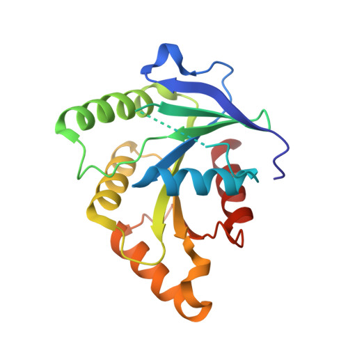
Analysis of the Open and Closed Conformations of the GTP-binding Protein YsxC from Bacillus subtilis.
Ruzheinikov, S.N., Das, S.K., Sedelnikova, S.E., Baker, P.J., Artymiuk, P.J., Garcia-Lara, J., Foster, S.J., Rice, D.W.(2004) J Mol Biology 339: 265-278
- PubMed: 15136032
- DOI: https://doi.org/10.1016/j.jmb.2004.03.043
- Primary Citation of Related Structures:
1SUL, 1SVI, 1SVW - PubMed Abstract:
Genetic analysis has suggested that the product of the Bacillus subtilis ysxC gene is essential for survival of the microorganism and hence may represent a target for the development of a novel anti-infective agent. B.subtilis YsxC is a member of the translation factor related class of GTPases and its crystal structure has been determined in an apo form and in complex with GDP and GMPPNP/Mg2+. Analysis of these structures has allowed us to examine the conformational changes that occur during the process of nucleotide binding and GTP hydrolysis. These structural changes particularly affect parts of the switch I and switch II region of YsxC, which become ordered and disordered, respectively in the "closed" or "on" GTP-bound state and disordered and ordered, respectively, in the "open" or "off" GDP-bound conformation. Finally, the binding of the magnesium cation results in subtle shifts of residues in the G3 region, at the start of switch II, which serve to optimize the interaction with a key aspartic acid residue. The structural flexibility observed in YsxC is likely to contribute to the role of the protein, possibly allowing transduction of an essential intracellular signal, which may be mediated via interactions with a conserved patch of surface-exposed, basic residues that lies adjacent to the GTP-binding site.
- Department of Molecular Biology and Biotechnology, Krebs Institute for Biomolecular Research, University of Sheffield, Firth Court, Western Bank, Sheffield S10 2TN, UK.
Organizational Affiliation:


