Multiconformational NMR analysis of sandostatin (octreotide): equilibrium between beta-sheet and partially helical structures.
Melacini, G., Zhu, Q., Goodman, M.(1997) Biochemistry 36: 1233-1241
- PubMed: 9063871 Search on PubMed
- DOI: https://doi.org/10.1021/bi962497o
- Primary Citation Related Structures:
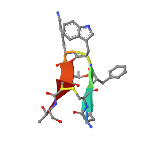
1SOC, 2SOC - PubMed Abstract:
This paper reports a detailed conformational analysis by 1H NMR (DMSO-d6, 300 K) and molecular modeling of the octapeptide D-Phe1-Cys2-Phe3-D-Trp4-Lys5-Thr6-Cys7+ ++-Thr8-ol (disulfide bridged) known as sandostatin (or SMS 201-995 or octreotide) with both somatostatin-like and opioid-like bioactivities. This is the initial report on sandostatin showing that attempts to explain all NMR data using a single average conformation reveal several important inconsistencies including severe violations of mutually exclusive backbone-to-backbone NOEs. The inconsistencies are solved by assuming an equilibrium between antiparallel beta-sheet structures and conformations in which the C-terminal residues form a 3(10) helix-like fold (helical ensemble). This conformational equilibrium is consistent with previous X-ray diffraction investigations which show that sandostatin can adopt both the beta-sheet and the 3(10) helix-like secondary structure folds. In addition, indications of a conformational equilibrium between beta-sheet and helical structures are also found in solvent systems different from DMSO-d6 and for other highly bioactive analogs of sandostatin. In these cases a proper multiconformational NMR refinement is important in order to avoid conformational averaging artifacts. Finally, using the known models for somatostatin-like and opioid-like bioactivities of sandostatin analogs, the present investigation shows the potentials of the proposed structures for the design of novel sandostatin-based conformationally restricted peptidomimetics. These analogs are expected to refine the pharmacophore models for sandostatin bioactivities.
- Department of Chemistry and Biochemistry, University of California at San Diego, La Jolla 92093-0343, USA.
Organizational Affiliation: