1SKS | pdb_00001sks

Binary 3' complex of T7 DNA polymerase with a DNA primer/template containing a cis-syn thymine dimer on the template


Experimental Data Snapshot

  • Method: X-RAY DIFFRACTION
  • Resolution: 2.30 Å
  • R-Value Free: 
    0.266 (Depositor), 0.272 (DCC) 
  • R-Value Work: 
    0.235 (Depositor), 0.244 (DCC) 
  • R-Value Observed: 
    0.235 (Depositor) 

wwPDB Validation 3D Report Full Report

Validation slider image for 1SKS

This is version 1.3 of the entry. See complete history

Literature

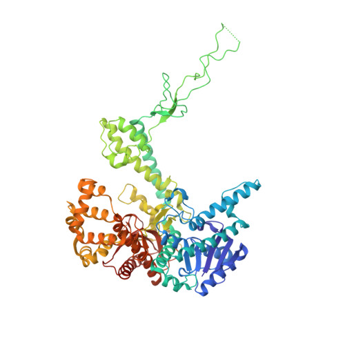
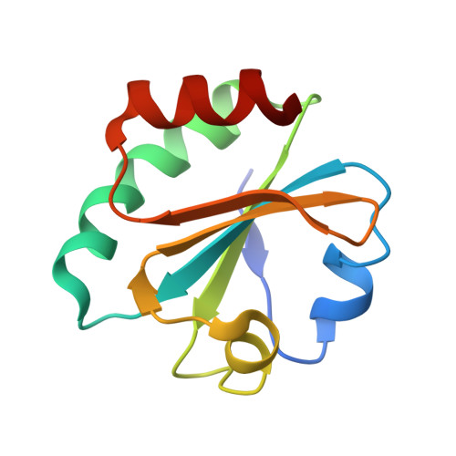
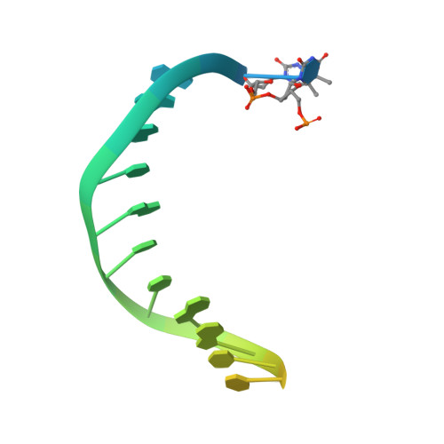
Nucleotide insertion opposite a cis-syn thymine dimer by a replicative DNA polymerase from bacteriophage T7.

Li, Y.Dutta, S.Doublie, S.Bdour, H.M.Taylor, J.S.Ellenberger, T.

(2004) Nat Struct Mol Biol 11: 784-790

  • DOI: https://doi.org/10.1038/nsmb792
  • Primary Citation Related Structures: 
    1SKR, 1SKS, 1SKW, 1SL0, 1SL1, 1SL2

  • PubMed Abstract: 

    Ultraviolet-induced DNA damage poses a lethal block to replication. To understand the structural basis for this, we determined crystal structures of a replicative DNA polymerase from bacteriophage T7 in complex with nucleotide substrates and a DNA template containing a cis-syn cyclobutane pyrimidine dimer (CPD). When the 3' thymine is the templating base, the CPD is rotated out of the polymerase active site and the fingers subdomain adopts an open orientation. When the 5' thymine is the templating base, the CPD lies within the polymerase active site where it base-pairs with the incoming nucleotide and the 3' base of the primer, while the fingers are in a closed conformation. These structures reveal the basis for the strong block of DNA replication that is caused by this photolesion.


  • Organizational Affiliation
    • Department of Biological Chemistry and Molecular Pharmacology, Harvard Medical School, 240 Longwood Avenue, Boston, Massachusetts 02115, USA.

Macromolecule Content 

  • Total Structure Weight: 105.15 kDa 
  • Atom Count: 6,769 
  • Modeled Residue Count: 809 
  • Deposited Residue Count: 852 
  • Unique protein chains: 2
  • Unique nucleic acid chains: 2

Macromolecules


Find similar proteins by:|  3D Structure
Entity ID: 3
MoleculeChains  Sequence LengthOrganismDetailsImage
DNA polymeraseC [auth A]698Escherichia phage T7Mutation(s): 0 
Gene Names: 5
EC: 2.7.7.7 (PDB Primary Data), 3.1.11 (UniProt)
UniProt
Find proteins for P00581 (Escherichia phage T7)
Explore P00581 
Go to UniProtKB:  P00581
Entity Groups
Sequence Clusters30% Identity50% Identity70% Identity90% Identity95% Identity100% Identity
UniProt GroupP00581
Sequence Annotations
Expand
Reference Sequence
Find similar proteins by:|  3D Structure
Entity ID: 4
MoleculeChains  Sequence LengthOrganismDetailsImage
Thioredoxin 1D [auth B]108Escherichia coliMutation(s): 0 
Gene Names: 
UniProt
Find proteins for P0AA25 (Escherichia coli (strain K12))
Explore P0AA25 
Go to UniProtKB:  P0AA25
Entity Groups
Sequence Clusters30% Identity50% Identity70% Identity90% Identity95% Identity100% Identity
UniProt GroupP0AA25
Sequence Annotations
Expand
Reference Sequence
Find similar nucleic acids by:  Sequence
Entity ID: 1
MoleculeChains LengthOrganismImage
5'-D(*CP*GP*AP*AP*AP*AP*CP*GP*AP*C*GP*GP*CP*CP*AP*GP*TP*GP*CP*CP*(2DT))-3'A [auth P]21N/A
Sequence Annotations
Expand
Reference Sequence
Find similar nucleic acids by:  Sequence
Entity ID: 2
MoleculeChains LengthOrganismImage
5'-D(*CP*CP*CP*(TTD)P*AP*GP*GP*CP*AP*CP*TP*GP*GP*CP*CP*GP*TP*CP*GP*TP*TP*TP*TP*CP*G)-3'B [auth T]25N/A
Sequence Annotations
Expand
Reference Sequence

Small Molecules

Ligands 1 Unique
IDChains Name / Formula / InChI Key2D Diagram3D Interactions
MG

Query on MG


Download:Ideal Coordinates CCD File
E [auth A]MAGNESIUM ION
Mg
JLVVSXFLKOJNIY-UHFFFAOYSA-N

Experimental Data & Validation

Experimental Data

  • Method: X-RAY DIFFRACTION
  • Resolution: 2.30 Å
  • R-Value Free:  0.266 (Depositor), 0.272 (DCC) 
  • R-Value Work:  0.235 (Depositor), 0.244 (DCC) 
  • R-Value Observed: 0.235 (Depositor) 
Space Group: P 21 21 2
Unit Cell:
Length ( Å )Angle ( ˚ )
a = 104.773α = 90
b = 214.917β = 90
c = 52.257γ = 90
Software Package:
Software NamePurpose
CNSrefinement
HKL-2000data reduction
SCALEPACKdata scaling
EPMRphasing

Structure Validation

View Full Validation Report


Entry History 

Deposition Data

Revision History  (Full details and data files)

  • Version 1.0: 2004-07-06
    Type: Initial release
  • Version 1.1: 2008-04-29
    Changes: Version format compliance
  • Version 1.2: 2011-07-13
    Changes: Version format compliance
  • Version 1.3: 2024-02-14
    Changes: Data collection, Database references, Derived calculations, Structure summary