Structural and functional basis of the serine protease-like hepatocyte growth factor beta-chain in Met binding and signaling
Kirchhofer, D., Yao, X., Peek, M., Eigenbrot, C., Lipari, M.T., Billeci, K.L., Maun, H.R., Moran, P., Santell, L., Wiesmann, C., Lazarus, R.A.(2004) J Biological Chem 279: 39915-39924
- PubMed: 15218027 Search on PubMed
- DOI: https://doi.org/10.1074/jbc.M404795200
- Primary Citation Related Structures:
1SI5 - PubMed Abstract:
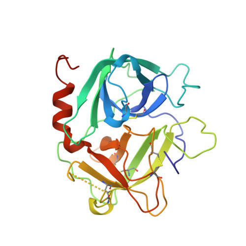
Hepatocyte growth factor (HGF), a plasminogen-related growth factor, is the ligand for Met, a receptor tyrosine kinase implicated in development, tissue regeneration, and invasive tumor growth. HGF acquires signaling activity only upon proteolytic cleavage of single-chain HGF into its alpha/beta heterodimer, similar to zymogen activation of structurally related serine proteases. Although both chains are required for activation, only the alpha-chain binds Met with high affinity. Recently, we reported that the protease-like HGF beta-chain binds to Met with low affinity (Stamos, J., Lazarus, R. A., Yao, X., Kirchhofer, D., and Wiesmann, C. (2004) EMBO J. 23, 2325-2335). Here we demonstrate that the zymogen-like form of HGF beta also binds Met, albeit with 14-fold lower affinity than the protease-like form, suggesting optimal interactions result from conformational changes upon cleavage of the single-chain form. Extensive mutagenesis of the HGF beta region corresponding to the active site and activation domain of serine proteases showed that 17 of the 38 purified two-chain HGF mutants resulted in impaired cell migration or Met phosphorylation but no loss in Met binding. However, reduced biological activities were well correlated with reduced Met binding of corresponding mutants of HGF beta itself in assays eliminating dominant alpha-chain binding contributions. Moreover, the crystal structure of HGF beta determined at 2.53 A resolution provides a structural context for the mutagenesis data. The functional Met binding site is centered on the "active site region" including "triad" residues Gln(534) [c57], Asp(578) [c102], and Tyr(673) [c195] and neighboring "activation domain" residues Val(692), Pro(693), Gly(694), Arg(695), and Gly(696) [c214-c219]. Together they define a region that bears remarkable resemblance to substrate processing regions of serine proteases. Models of HGF-dependent Met receptor activation are discussed.
- Department of Physiology, Genentech, Inc., South San Francisco, California 94080, USA. dak@gene.com
Organizational Affiliation: