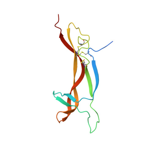
1SGF

CRYSTAL STRUCTURE OF 7S NGF: A COMPLEX OF NERVE GROWTH FACTOR WITH FOUR BINDING PROTEINS (SERINE PROTEINASES)


Experimental Data Snapshot

  • Method: X-RAY DIFFRACTION
  • Resolution: 3.15 Å
  • R-Value Free: 0.282 
  • R-Value Work: 0.246 
  • R-Value Observed: 0.246 

Starting Models: experimental
View more details

wwPDB Validation   3D Report Full Report


Ligand Structure Quality Assessment 


This is version 2.1 of the entry. See complete history


Literature

Structure of mouse 7S NGF: a complex of nerve growth factor with four binding proteins.

Bax, B.Blundell, T.L.Murray-Rust, J.McDonald, N.Q.

(1997) Structure 5: 1275-1285

  • DOI: https://doi.org/10.1016/s0969-2126(97)00280-3
  • Primary Citation of Related Structures:  
    1SGF

  • PubMed Abstract: 

    Nerve growth factor (NGF) is a neurotrophic factor that promotes the differentiation and survival of certain populations of neurons in the central and peripheral nervous systems. 7S NGF is an alpha 2 beta 2 gamma 2 complex in which the beta-NGF dimer (the active neurotrophin) is associated with two alpha-NGF and two gamma-NGF subunits, which belong to the glandular kallikrein family of serine proteinases. The gamma-NGF subunit is an active serine proteinase capable of processing the precursor form of beta-NGF, whereas alpha-NGF is an inactive serine proteinase. The structure of 7S NGF could be used as a starting point to design inhibitors that prevent NGF binding to its receptors, as a potential treatment of neurodegenerative diseases. The crystal structure of 7S NGF shows that the two gamma-NGF subunits make extensive interactions with each other around the twofold axis of the complex and have the C-terminal residues of the beta-NGF subunits bound within their active sites. The 'activation domain' of each of the alpha-NGF subunits is in an inactive (zymogen-like) conformation and makes extensive interactions with the beta-NGF dimer. The two zinc ions that stabilize the complex are located at the relatively small interfaces between the alpha-NGF and gamma-NGF subunits. The structure of 7S NGF shows how the twofold axis of the central beta-NGF dimer organizes the symmetry of this multisubunit growth factor complex. The extensive surface of beta-NGF buried within the 7S complex explains the lack of neurotrophic activity observed for 7S NGF. The regions of the beta-NGF dimer that contact the alpha-NGF subunits overlap with those known to engage NGF receptors. Two disulphide-linked loops on alpha-NGF make multiple interactions with beta-NGF and suggest that it might be possible to design peptides that inhibit the binding of beta-NGF to its receptors.


  • Organizational Affiliation

    Department of Crystallography, Birkbeck College, London, UK. b.bax@cryst.bbk.ac.uk


Macromolecules
Find similar proteins by:  (by identity cutoff)  |  3D Structure
Entity ID: 1
MoleculeChains Sequence LengthOrganismDetailsImage
NERVE GROWTH FACTORA,
D [auth X]
240Mus musculusMutation(s): 0 
EC: 3.4.21.35
UniProt
Find proteins for P00757 (Mus musculus)
Explore P00757 
Go to UniProtKB:  P00757
Entity Groups  
Sequence Clusters30% Identity50% Identity70% Identity90% Identity95% Identity100% Identity
UniProt GroupP00757
Glycosylation
Glycosylation Sites: 1Go to GlyGen: P00757-1
Sequence Annotations
Expand
  • Reference Sequence
Find similar proteins by:  (by identity cutoff)  |  3D Structure
Entity ID: 2
MoleculeChains Sequence LengthOrganismDetailsImage
NERVE GROWTH FACTORB,
E [auth Y]
118Mus musculusMutation(s): 0 
EC: 3.4.21.35
UniProt
Find proteins for P01139 (Mus musculus)
Explore P01139 
Go to UniProtKB:  P01139
Entity Groups  
Sequence Clusters30% Identity50% Identity70% Identity90% Identity95% Identity100% Identity
UniProt GroupP01139
Sequence Annotations
Expand
  • Reference Sequence
Find similar proteins by:  (by identity cutoff)  |  3D Structure
Entity ID: 3
MoleculeChains Sequence LengthOrganismDetailsImage
NERVE GROWTH FACTORC [auth G],
F [auth Z]
237Mus musculusMutation(s): 0 
EC: 3.4.21.35
UniProt
Find proteins for P00756 (Mus musculus)
Explore P00756 
Go to UniProtKB:  P00756
Entity Groups  
Sequence Clusters30% Identity50% Identity70% Identity90% Identity95% Identity100% Identity
UniProt GroupP00756
Glycosylation
Glycosylation Sites: 1Go to GlyGen: P00756-1
Sequence Annotations
Expand
  • Reference Sequence
Oligosaccharides

Help

Entity ID: 4
MoleculeChains Length2D Diagram Glycosylation3D Interactions
2-acetamido-2-deoxy-beta-D-glucopyranose-(1-4)-2-acetamido-2-deoxy-beta-D-glucopyranoseG [auth C],
H [auth D]
2N-Glycosylation
Glycosylation Resources
GlyTouCan:  G42666HT
GlyCosmos:  G42666HT
GlyGen:  G42666HT
Experimental Data & Validation

Experimental Data

  • Method: X-RAY DIFFRACTION
  • Resolution: 3.15 Å
  • R-Value Free: 0.282 
  • R-Value Work: 0.246 
  • R-Value Observed: 0.246 
  • Space Group: P 21 21 21
Unit Cell:
Length ( Å )Angle ( ˚ )
a = 95.68α = 90
b = 96.59β = 90
c = 147γ = 90
Software Package:
Software NamePurpose
MOSFLMdata reduction
ROTAVATA/AGROVATAdata reduction
TFFCmodel building
X-PLORmodel building
X-PLORrefinement
CCP4data scaling
ROTAVATAdata scaling
TFFCphasing
X-PLORphasing

Structure Validation

View Full Validation Report


Ligand Structure Quality Assessment 


Entry History 

Deposition Data

Revision History  (Full details and data files)

  • Version 1.0: 1998-05-27
    Type: Initial release
  • Version 1.1: 2008-03-24
    Changes: Version format compliance
  • Version 1.2: 2011-07-13
    Changes: Non-polymer description, Version format compliance
  • Version 2.0: 2020-07-29
    Type: Remediation
    Reason: Carbohydrate remediation
    Changes: Advisory, Atomic model, Data collection, Derived calculations, Non-polymer description, Refinement description, Structure summary
  • Version 2.1: 2023-08-09
    Changes: Database references, Refinement description, Structure summary