1SF6

BINDING OF N,N',N"-TRIACETYLCHITOTRIOSE TO HEW LYSOZYME: A POWDER DIFFRACTION STUDY


Experimental Data Snapshot

  • Method: POWDER DIFFRACTION

wwPDB Validation   3D Report Full Report


This is version 2.0 of the entry. See complete history


Literature

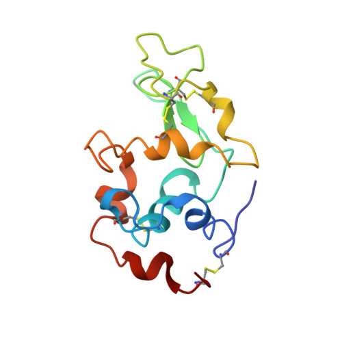
Binding of N-acetylglucosamine oligosaccharides to hen egg-white lysozyme: a powder diffraction study.

Von Dreele, R.B.

(2005) Acta Crystallogr D Biol Crystallogr 61: 22-32

  • DOI: https://doi.org/10.1107/S0907444904025715
  • Primary Citation of Related Structures:  
    1SF4, 1SF6, 1SF7, 1SFB, 1SFG

  • PubMed Abstract: 

    The binding of N-acetylglucosamine oligosaccharides (NAGn, n = 2-6) to hen egg-white lysozyme (HEWL; EC 3.2.1.17) was investigated by X-ray powder diffraction at room temperature. Each NAGn examined was found to bind to lysozyme in rapid-precipitation preparations in 1.0 M NaCl pH 6.0 buffer. The location of each NAGn was easily found from difference Fourier maps generated from structure factors extracted during preliminary Rietveld refinements. Full NAGn-protein structures were subjected to combined Rietveld and stereochemical restraint refinements (Rwp = 2.28-2.59%; Rp = 1.81-2.04%; RF2 = 3.91-5.80%) and revealed binding modes for NAGn that depended on the length of the NAG oligosaccharide. The NAG2 ligand was found in the BC sites in the cleft of HEWL, NAG3 was found to bind in both the ABC and BCD sites in the ratio 35:65 and NAG4 and NAG5 bound to the ABCD and ABCDE sites, respectively, while NAG6 only bound to sites ABCDE, leaving the F site empty with the remaining saccharide ring located in a solvent region adjacent to the A site. All protein powder diffraction patterns in this study consisted of extremely sharp Bragg peaks consistent with approximately 1 microm crystallites that were devoid of line-broadening defects. Details of the stereochemical restraints used in these refinements and their impact on structural validation are also discussed.


  • Organizational Affiliation

    XPS/IPNS Divisions, Argonne National Laboratory, Argonne, IL 60439, USA. vondreele@anl.gov


Macromolecules
Find similar proteins by:  (by identity cutoff)  |  3D Structure
Entity ID: 1
MoleculeChains Sequence LengthOrganismDetailsImage
LYSOZYME129Gallus gallusMutation(s): 0 
EC: 3.2.1.17
UniProt
Find proteins for P00698 (Gallus gallus)
Explore P00698 
Go to UniProtKB:  P00698
Entity Groups  
Sequence Clusters30% Identity50% Identity70% Identity90% Identity95% Identity100% Identity
UniProt GroupP00698
Sequence Annotations
Expand
  • Reference Sequence
Oligosaccharides

Help

Entity ID: 2
MoleculeChains Length2D Diagram Glycosylation3D Interactions
2-acetamido-2-deoxy-beta-D-glucopyranose-(1-4)-2-acetamido-2-deoxy-beta-D-glucopyranose-(1-4)-2-acetamido-2-deoxy-beta-D-glucopyranose-(1-4)-2-acetamido-2-deoxy-beta-D-glucopyranose
B
4N/A
Glycosylation Resources
GlyTouCan:  G01361SX
GlyCosmos:  G01361SX
GlyGen:  G01361SX
Experimental Data & Validation

Experimental Data

  • Method: POWDER DIFFRACTION
  • Space Group: P 43 21 2
Unit Cell:
Length ( Å )Angle ( ˚ )
a = 78.77α = 90
b = 78.77β = 90
c = 38.368γ = 90
Software Package:
Software NamePurpose
povdata collection
PROCESSdata reduction
GSASmodel building
GSASrefinement
POVdata reduction
PROCESSdata scaling
GSASphasing

Structure Validation

View Full Validation Report


Entry History 

Deposition Data

Revision History  (Full details and data files)

  • Version 1.0: 2004-03-02
    Type: Initial release
  • Version 1.1: 2008-04-29
    Changes: Version format compliance
  • Version 1.2: 2011-07-13
    Changes: Non-polymer description, Version format compliance
  • Version 2.0: 2020-07-29
    Type: Remediation
    Reason: Carbohydrate remediation
    Changes: Advisory, Atomic model, Data collection, Derived calculations, Structure summary