1S1R | pdb_00001s1r

Crystal structures of prostaglandin D2 11-ketoreductase (AKR1C3) in complex with the non-steroidal anti-inflammatory drugs flufenamic acid and indomethacin


Experimental Data Snapshot

  • Method: X-RAY DIFFRACTION
  • Resolution: 2.00 Å
  • R-Value Free: 
    0.196 (Depositor) 
  • R-Value Work: 
    0.158 (Depositor) 
  • R-Value Observed: 
    0.160 (Depositor) 

Starting Model: experimental
View more details

wwPDB Validation 3D Report Full Report

Validation slider image for 1S1R

This is version 1.3 of the entry. See complete history

Literature

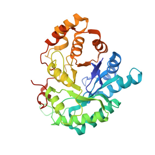
Crystal structures of prostaglandin D(2) 11-ketoreductase (AKR1C3) in complex with the nonsteroidal anti-inflammatory drugs flufenamic acid and indomethacin.

Lovering, A.L.Ride, J.P.Bunce, C.M.Desmond, J.C.Cummings, S.M.White, S.A.

(2004) Cancer Res 64: 1802-1810

  • DOI: https://doi.org/10.1158/0008-5472.can-03-2847
  • Primary Citation Related Structures: 
    1S1P, 1S1R, 1S2A, 1S2C

  • PubMed Abstract: 

    It is becoming increasingly well established that nonsteroidal anti-inflammatory drugs (NSAID) protect against tumors of the gastrointestinal tract and that they may also protect against a variety of other tumors. These activities have been widely attributed to the inhibition of cylooxygenases (COX) and, in particular, COX-2. However, several observations have indicated that other targets may be involved. Besides targeting COX, certain NSAID also inhibit enzymes belonging to the aldo-keto reductase (AKR) family, including AKR1C3. We have demonstrated previously that overexpression of AKR1C3 acts to suppress cell differentiation and promote proliferation in myeloid cells. However, this enzyme has a broad tissue distribution and therefore represents a novel candidate for the target of the COX-independent antineoplastic actions of NSAID. Here we report on the X-ray crystal structures of AKR1C3 complexed with the NSAID indomethacin (1.8 A resolution) or flufenamic acid (1.7 A resolution). One molecule of indomethacin is bound in the active site, whereas flufenamic acid binds to both the active site and the beta-hairpin loop, at the opposite end of the central beta-barrel. Two other crystal structures (1.20 and 2.1 A resolution) show acetate bound in the active site occupying the proposed oxyanion hole. The data underline AKR1C3 as a COX-independent target for NSAID and will provide a structural basis for the future development of new cancer therapies with reduced COX-dependent side effects.


  • Organizational Affiliation
    • The School of Biosciences, The University of Birmingham, Birmingham, United Kingdom.

Macromolecule Content 

  • Total Structure Weight: 38.89 kDa 
  • Atom Count: 2,888 
  • Modeled Residue Count: 316 
  • Deposited Residue Count: 331 
  • Unique protein chains: 1

Macromolecules

Find similar proteins by:|  3D Structure
Entity ID: 1
MoleculeChains  Sequence LengthOrganismDetailsImage
Aldo-keto reductase family 1 member C3331Homo sapiensMutation(s): 0 
EC: 1.1.1.213 (PDB Primary Data), 1.3.1.20 (PDB Primary Data), 1.1.1.62 (PDB Primary Data), 1.1.1.64 (UniProt), 1.1.1.53 (UniProt), 1.1.1.357 (UniProt), 1.1.1.188 (UniProt), 1.1.1.239 (UniProt), 1.1.1 (UniProt), 1.1.1.210 (UniProt)
UniProt & NIH Common Fund Data Resources
Find proteins for P42330 (Homo sapiens)
Explore P42330 
Go to UniProtKB:  P42330
PHAROS:  P42330
GTEx:  ENSG00000196139 
Entity Groups
Sequence Clusters30% Identity50% Identity70% Identity90% Identity95% Identity100% Identity
UniProt GroupP42330
Sequence Annotations
Expand
Reference Sequence

Small Molecules

Ligands 3 Unique
IDChains Name / Formula / InChI Key2D Diagram3D Interactions
NAP

Query on NAP


Download:Ideal Coordinates CCD File
C [auth A]NADP NICOTINAMIDE-ADENINE-DINUCLEOTIDE PHOSPHATE
C21 H28 N7 O17 P3
XJLXINKUBYWONI-NNYOXOHSSA-N
MPD

Query on MPD


Download:Ideal Coordinates CCD File
D [auth A](4S)-2-METHYL-2,4-PENTANEDIOL
C6 H14 O2
SVTBMSDMJJWYQN-YFKPBYRVSA-N
ACT

Query on ACT


Download:Ideal Coordinates CCD File
B [auth A]ACETATE ION
C2 H3 O2
QTBSBXVTEAMEQO-UHFFFAOYSA-M

Experimental Data & Validation

Experimental Data

  • Method: X-RAY DIFFRACTION
  • Resolution: 2.00 Å
  • R-Value Free:  0.196 (Depositor) 
  • R-Value Work:  0.158 (Depositor) 
  • R-Value Observed: 0.160 (Depositor) 
Space Group: P 32 2 1
Unit Cell:
Length ( Å )Angle ( ˚ )
a = 57.917α = 90
b = 57.917β = 90
c = 206.168γ = 120
Software Package:
Software NamePurpose
REFMACrefinement
MOSFLMdata reduction
CCP4data scaling
CNSphasing

Structure Validation

View Full Validation Report


Entry History 

Deposition Data

Revision History  (Full details and data files)

  • Version 1.0: 2004-03-23
    Type: Initial release
  • Version 1.1: 2008-04-29
    Changes: Version format compliance
  • Version 1.2: 2011-07-13
    Changes: Version format compliance
  • Version 1.3: 2023-10-25
    Changes: Data collection, Database references, Derived calculations, Refinement description