1RZD | pdb_00001rzd

X-RAY ANALYSIS OF METAL SUBSTITUTED HUMAN CARBONIC ANHYDRASE II DERIVATIVES


Experimental Data Snapshot

  • Method: X-RAY DIFFRACTION
  • Resolution: 1.90 Å
  • R-Value Work: 
    0.141 (DCC) 
  • R-Value Observed: 
    0.148 (Depositor) 

wwPDB Validation 3D Report Full Report

Validation slider image for 1RZD

This is version 1.4 of the entry. See complete history

Literature

X-ray analysis of metal-substituted human carbonic anhydrase II derivatives.

Hakansson, K.Wehnert, A.Liljas, A.

(1994) Acta Crystallogr D Biol Crystallogr 50: 93-100

  • DOI: https://doi.org/10.1107/S0907444993008790
  • Primary Citation Related Structures: 
    1RZA, 1RZB, 1RZC, 1RZD, 1RZE

  • PubMed Abstract: 

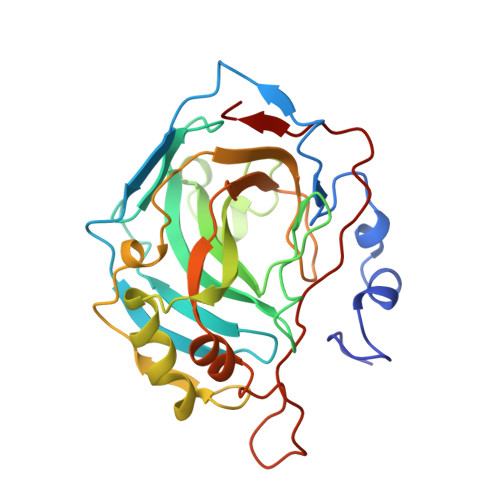
    Metal-substituted crystals of human carbonic anhydrase II belonging to space group P2(1) with cell dimensions a = 42.7, b = 41.7, c = 73.0 A and beta = 104.6 degrees were analyzed crystallographically. The resolution limit ranged from 1.82 to 1.92 A with high completeness (86.2-90.7%). Cobalt(II)-substituted carbonic anhydrase has a tetrahedral coordination around the metal both at pH 6 and pH 7.8, similar to the native zinc enzyme. In contrast, the catalytically inactive copper(II), nickel(II) and manganese(II) derivatives showed increased coordination number around the metal ion. Whereas the copper is best described as penta-coordinated, the nickel and manganese are best described as hexa-coordinated. The results are briefly compared with spectroscopic observations and our current view on carbonic anhydrase catalysis.


  • Organizational Affiliation
    • Molecular Biophysics, Chemical Center, University of Lund, Sweden.

Macromolecule Content 

  • Total Structure Weight: 29.31 kDa 
  • Atom Count: 2,301 
  • Modeled Residue Count: 258 
  • Deposited Residue Count: 259 
  • Unique protein chains: 1

Macromolecules

Find similar proteins by:|  3D Structure
Entity ID: 1
MoleculeChains  Sequence LengthOrganismDetailsImage
CARBONIC ANHYDRASE II259Homo sapiensMutation(s): 0 
EC: 4.2.1.1 (PDB Primary Data), 4.2.1.69 (UniProt)
UniProt & NIH Common Fund Data Resources
Find proteins for P00918 (Homo sapiens)
Explore P00918 
Go to UniProtKB:  P00918
PHAROS:  P00918
GTEx:  ENSG00000104267 
Entity Groups
Sequence Clusters30% Identity50% Identity70% Identity90% Identity95% Identity100% Identity
UniProt GroupP00918
Sequence Annotations
Expand
Reference Sequence

Experimental Data & Validation

Experimental Data

  • Method: X-RAY DIFFRACTION
  • Resolution: 1.90 Å
  • R-Value Work:  0.141 (DCC) 
  • R-Value Observed: 0.148 (Depositor) 
Space Group: P 1 21 1
Unit Cell:
Length ( Å )Angle ( ˚ )
a = 42.7α = 90
b = 41.7β = 104.6
c = 73γ = 90
Software Package:
Software NamePurpose
PROFFTrefinement

Structure Validation

View Full Validation Report


Entry History 

Deposition Data

Revision History  (Full details and data files)

  • Version 1.0: 1993-10-31
    Type: Initial release
  • Version 1.1: 2008-03-03
    Changes: Version format compliance
  • Version 1.2: 2011-07-13
    Changes: Version format compliance
  • Version 1.3: 2017-11-29
    Changes: Derived calculations, Other
  • Version 1.4: 2024-02-14
    Changes: Data collection, Database references, Derived calculations