1RJU | pdb_00001rju

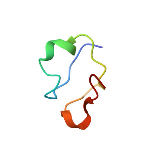
Crystal structure of a truncated form of yeast copper thionein


Experimental Data Snapshot

  • Method: X-RAY DIFFRACTION
  • Resolution: 1.44 Å
  • R-Value Free: 
    0.171 (Depositor) 
  • R-Value Work: 
    0.145 (Depositor), 0.150 (DCC) 
  • R-Value Observed: 
    0.147 (Depositor) 

wwPDB Validation 3D Report Full Report

Validation slider image for 1RJU

This is version 1.3 of the entry. See complete history

Literature

The Crystal Structure of Yeast Copper Thionein: the solution of a long lasting Enigma.

Calderone, V.Dolderer, B.Hartmann, H.J.Echner, H.Luchinat, C.Del Bianco, C.Mangani, S.Weser, U.

(2005) Proc Natl Acad Sci U S A 102: 51-56

  • DOI: https://doi.org/10.1073/pnas.0408254101
  • Primary Citation Related Structures: 
    1RJU

  • PubMed Abstract: 

    We report here the crystal structure of yeast copper thionein (Cu-MT), determined at 1.44-A resolution. The Cu-MT structure shows the largest known oligonuclear Cu(I) thiolate cluster in biology, consisting of six trigonally and two digonally coordinated Cu(I) ions. This is at variance with the results from previous spectroscopic determinations, which were performed on MT samples containing seven rather than eight metal ions. The protein backbone has a random coil structure with the loops enfolding the copper cluster, which is located in a cleft where it is bound to 10 cysteine residues. The protein structure is somewhat different from that of Ag(7)-MT and similar, but not identical, to that of Cu(7)-MT. Besides the different structure of the metal cluster, the main differences lie in the cysteine topology and in the conformation of some portions of the backbone. The present structure suggests that Cu-MT, in addition to its role as a safe depository for copper ions in the cell, may play an active role in the delivery of copper to metal-free chaperones.


  • Organizational Affiliation
    • Department of Chemistry, University of Siena, Via Aldo Moro, 53100 Siena, Italy.

Macromolecule Content 

  • Total Structure Weight: 4.36 kDa 
  • Atom Count: 302 
  • Modeled Residue Count: 36 
  • Deposited Residue Count: 36 
  • Unique protein chains: 1

Macromolecules

Find similar proteins by:|  3D Structure
Entity ID: 1
MoleculeChains  Sequence LengthOrganismDetailsImage
MetallothioneinA [auth V]36N/AMutation(s): 0 
UniProt
Find proteins for P0CX80 (Saccharomyces cerevisiae (strain ATCC 204508 / S288c))
Explore P0CX80 
Go to UniProtKB:  P0CX80
Entity Groups
Sequence Clusters30% Identity50% Identity70% Identity90% Identity95% Identity100% Identity
UniProt GroupP0CX80
Sequence Annotations
Expand
Reference Sequence

Experimental Data & Validation

Experimental Data

  • Method: X-RAY DIFFRACTION
  • Resolution: 1.44 Å
  • R-Value Free:  0.171 (Depositor) 
  • R-Value Work:  0.145 (Depositor), 0.150 (DCC) 
  • R-Value Observed: 0.147 (Depositor) 
Space Group: P 43 3 2
Unit Cell:
Length ( Å )Angle ( ˚ )
a = 62.167α = 90
b = 62.167β = 90
c = 62.167γ = 90
Software Package:
Software NamePurpose
REFMACrefinement
MOSFLMdata reduction
CCP4data scaling
SOLVEphasing
RESOLVEphasing

Structure Validation

View Full Validation Report


Entry History 

Deposition Data

Revision History  (Full details and data files)

  • Version 1.0: 2004-12-07
    Type: Initial release
  • Version 1.1: 2008-04-29
    Changes: Version format compliance
  • Version 1.2: 2011-07-13
    Changes: Version format compliance
  • Version 1.3: 2024-02-14
    Changes: Data collection, Database references, Derived calculations