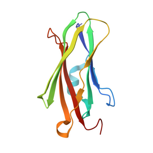
The impact of Lys-->Arg surface mutations on the crystallization of the globular domain of RhoGDI.
Czepas, J., Devedjiev, Y., Krowarsch, D., Derewenda, U., Otlewski, J., Derewenda, Z.S.(2004) Acta Crystallogr D Biol Crystallogr 60: 275-280
- PubMed: 14747703
- DOI: https://doi.org/10.1107/S0907444903026271
- Primary Citation of Related Structures:
1QVY - PubMed Abstract:
The potential of rational surface mutagenesis for enhanced protein crystallization is being probed in an ongoing effort. In previous work, it was hypothesized that residues with high conformational entropy such as Glu and Lys are suitable targets for surface mutagenesis, as they are rarely incorporated in crystal contacts or protein-protein interfaces. Previous experiments using Lys-->Ala, Glu-->Ala and Glu-->Asp mutants confirmed that mutated proteins were more likely to crystallize. In the present paper, the usefulness of Lys-->Arg mutations is studied. Several mutations of the globular domain of human RhoGDI were generated, including the single mutants K105R, K113R, K127R, K138R and K141R, the double mutants K(98,99)R and K(199,200)R and the triple mutants K(98,99,105)R and K(135,138,141)R. It is shown that Lys-->Arg mutants are more likely to crystallize than the wild-type protein, although not as likely as Lys-->Ala mutants. Out of the nine mutants tested, five produced diffracting crystals, including the K(199,200)R double mutant, which crystallized in a new space group and exceeded by approximately 1.0 A the resolution of the diffraction of the wild-type crystal. Major crystal contacts in the new lattice were created by the mutated epitope.
- Department of Molecular Physiology and Biological Physics, University of Virginia, Charlottesville, VA 22908-0736, USA.
Organizational Affiliation:


