1QUB

CRYSTAL STRUCTURE OF THE GLYCOSYLATED FIVE-DOMAIN HUMAN BETA2-GLYCOPROTEIN I PURIFIED FROM BLOOD PLASMA


Experimental Data Snapshot

  • Method: X-RAY DIFFRACTION
  • Resolution: 2.70 Å
  • R-Value Free: 0.269 
  • R-Value Work: 0.249 
  • R-Value Observed: 0.250 

wwPDB Validation   3D Report Full Report


This is version 2.0 of the entry. See complete history


Literature

Adhesion mechanism of human beta(2)-glycoprotein I to phospholipids based on its crystal structure.

Bouma, B.de Groot, P.G.van den Elsen, J.M.Ravelli, R.B.Schouten, A.Simmelink, M.J.Derksen, R.H.Kroon, J.Gros, P.

(1999) EMBO J 18: 5166-5174

  • DOI: https://doi.org/10.1093/emboj/18.19.5166
  • Primary Citation of Related Structures:  
    1QUB

  • PubMed Abstract: 

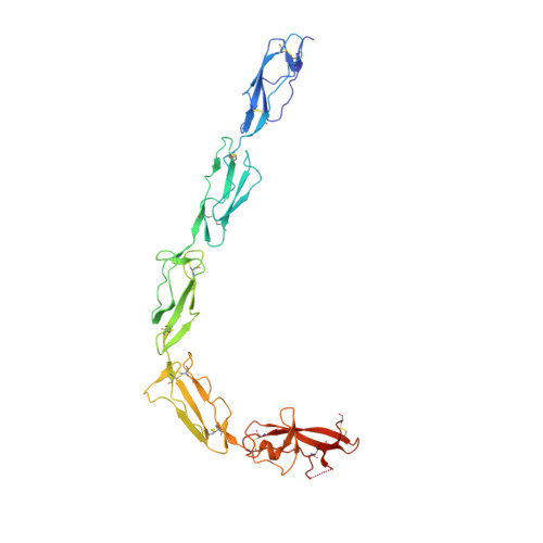
    Human beta(2)-glycoprotein I is a heavily glycosylated five-domain plasma membrane-adhesion protein, which has been implicated in blood coagulation and clearance of apoptotic bodies from the circulation. It is also the key antigen in the autoimmune disease anti-phospholipid syndrome. The crystal structure of beta(2)-glycoprotein I isolated from human plasma reveals an elongated fish-hook-like arrangement of the globular short consensus repeat domains. Half of the C-terminal fifth domain deviates strongly from the standard fold, as observed in domains one to four. This aberrant half forms a specific phospholipid-binding site. A large patch of 14 positively charged residues provides electrostatic interactions with anionic phospholipid headgroups and an exposed membrane-insertion loop yields specificity for lipid layers. The observed spatial arrangement of the five domains suggests a functional partitioning of protein adhesion and membrane adhesion over the N- and C-terminal domains, respectively, separated by glycosylated bridging domains. Coordinates are in the Protein Data Bank (accession No. 1QUB).


  • Organizational Affiliation

    Department of Crystal and Structural Chemistry, Bijvoet Center for Biomolecular Research, Utrecht University, Padualaan 8, 3584 CH Utrecht, The Netherlands.


Macromolecules
Find similar proteins by:  (by identity cutoff)  |  3D Structure
Entity ID: 1
MoleculeChains Sequence LengthOrganismDetailsImage
PROTEIN (human beta2-Glycoprotein I)319Homo sapiensMutation(s): 0 
Gene Names: APOHB2G1
UniProt & NIH Common Fund Data Resources
Find proteins for P02749 (Homo sapiens)
Explore P02749 
Go to UniProtKB:  P02749
PHAROS:  P02749
Entity Groups  
Sequence Clusters30% Identity50% Identity70% Identity90% Identity95% Identity100% Identity
UniProt GroupP02749
Sequence Annotations
Expand
  • Reference Sequence
Oligosaccharides

Help

Entity ID: 2
MoleculeChains Length2D Diagram Glycosylation3D Interactions
alpha-D-mannopyranose-(1-4)-2-acetamido-2-deoxy-beta-D-glucopyranose-(1-4)-2-acetamido-2-deoxy-beta-D-glucopyranose
B
3N-Glycosylation
Glycosylation Resources
GlyTouCan:  G62182OO
GlyCosmos:  G62182OO
GlyGen:  G62182OO
Entity ID: 3
MoleculeChains Length2D Diagram Glycosylation3D Interactions
2-acetamido-2-deoxy-beta-D-glucopyranose-(1-4)-2-acetamido-2-deoxy-beta-D-glucopyranose
C
2N-Glycosylation
Glycosylation Resources
GlyTouCan:  G42666HT
GlyCosmos:  G42666HT
GlyGen:  G42666HT
Small Molecules
Ligands 1 Unique
IDChains Name / Formula / InChI Key2D Diagram3D Interactions
NAG
Query on NAG

Download Ideal Coordinates CCD File 
D [auth A],
E [auth A]
2-acetamido-2-deoxy-beta-D-glucopyranose
C8 H15 N O6
OVRNDRQMDRJTHS-FMDGEEDCSA-N
Experimental Data & Validation

Experimental Data

  • Method: X-RAY DIFFRACTION
  • Resolution: 2.70 Å
  • R-Value Free: 0.269 
  • R-Value Work: 0.249 
  • R-Value Observed: 0.250 
  • Space Group: C 2 2 21
Unit Cell:
Length ( Å )Angle ( ˚ )
a = 161.17α = 90
b = 166.49β = 90
c = 114.51γ = 90
Software Package:
Software NamePurpose
MOSFLMdata reduction
TRUNCATEdata reduction
SOLVEphasing
CNSrefinement
CCP4data scaling

Structure Validation

View Full Validation Report


Entry History 

Deposition Data

Revision History  (Full details and data files)

  • Version 1.0: 1999-10-08
    Type: Initial release
  • Version 1.1: 2008-04-27
    Changes: Version format compliance
  • Version 1.2: 2011-07-13
    Changes: Non-polymer description, Version format compliance
  • Version 1.3: 2018-01-24
    Changes: Structure summary
  • Version 2.0: 2020-07-29
    Type: Remediation
    Reason: Carbohydrate remediation
    Changes: Advisory, Atomic model, Data collection, Derived calculations, Structure summary