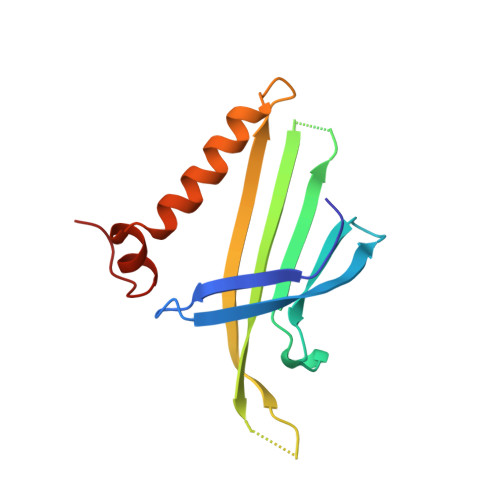
The crystal structure of bacteriophage Q beta at 3.5 A resolution.
Golmohammadi, R., Fridborg, K., Bundule, M., Valegard, K., Liljas, L.(1996) Structure 4: 543-554
- PubMed: 8736553
- DOI: https://doi.org/10.1016/s0969-2126(96)00060-3
- Primary Citation of Related Structures:
1QBE - PubMed Abstract:
The capsid protein subunits of small RNA bacteriophages form a T = 3 particle upon assembly and RNA encapsidation. Dimers of the capsid protein repress translation of the replicase gene product by binding to the ribosome binding site and this interaction is believed to initiate RNA encapsidation. We have determined the crystal structure of phage Q beta with the aim of clarifying which factors are the most important for particle assembly and RNA interaction in the small phages. The crystal structure of bacteriophage Q beta determined at 3.5 A resolution shows that the capsid is stabilized by disulfide bonds on each side of the flexible loops that are situated around the fivefold and quasi-sixfold axes. As in other small RNA phages, the protein capsid is constructed from subunits which associate into dimers. A contiguous ten-stranded antiparallel beta sheet facing the RNA is formed in the dimer. The disulfide bonds lock the constituent dimers of the capsid covalently in the T = 3 lattice. The unusual stability of the Q beta particle is due to the tight dimer interactions and the disulfide bonds linking each dimer covalently to the rest of the capsid. A comparison with the structure of the related phage MS2 shows that although the fold of the Q beta coat protein is very similar, the details of the protein-protein interactions are completely different. The most conserved region of the protein is at the surface, which, in MS2, is involved in RNA binding.
- Department of Molecular Biology, Uppsala University, BMC, Sweden.
Organizational Affiliation:


