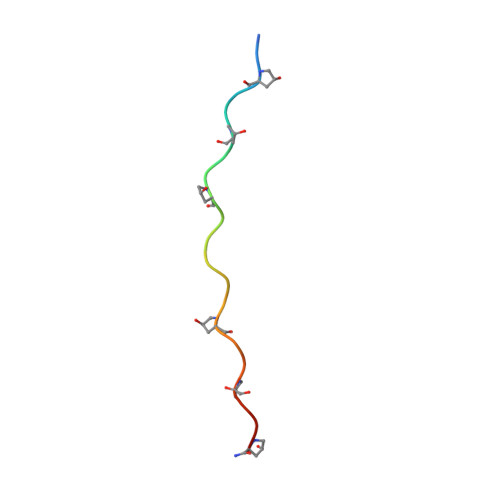
Structure of the Integrin alpha2beta1-binding Collagen Peptide.
Emsley, J., Knight, C.G., Farndale, R.W., Barnes, M.J.(2004) J Mol Biology 335: 1019-1028
- PubMed: 14698296 Search on PubMed
- DOI: https://doi.org/10.1016/j.jmb.2003.11.030
- Primary Citation Related Structures:
1Q7D - PubMed Abstract:
We have determined the 1.8A crystal structure of a triple helical integrin-binding collagen peptide (IBP) with sequence (Gly-Pro-Hyp)(2)-Gly-Phe-Hyp-Gly-Glu-Arg-(Gly-Pro-Hyp)(3). The central GFOGER hexapeptide is recognised specifically by the integrins alpha2beta1, alpha1beta1, alpha10beta1 and alpha11beta1. These integrin/collagen interactions are implicated in a number of key physiological processes including cell adhesion, cell growth and differentiation, and pathological states such as thrombosis and tumour metastasis. Comparison of the IBP structure with the previously determined structure of an identical collagen peptide in complex with the integrin alpha2-I domain (IBP(c)) allows the first detailed examination of collagen in a bound and an unbound state. The IBP structure shows a direct and a water-mediated electrostatic interaction between Glu and Arg side-chains from adjacent strands, but no intra-strand interactions. The interactions between IBP Glu and Arg side-chains are disrupted upon integrin binding. A comparison of IBP and IBP(c) main-chain conformation reveals the flexible nature of the triple helix backbone in the imino-poor GFOGER region. This flexibility could be important to the integrin-collagen interaction and provides a possible explanation for the unique orientation of the three GFOGER strands observed in the integrin-IBP(c) complex crystal structure.
- Department of Biochemistry, University of Leicester LE17RH, Leicester, UK. je14@le.ac.uk
Organizational Affiliation: