1Q1K

Structure of ATP-phosphoribosyltransferase from E. coli complexed with PR-ATP


Experimental Data Snapshot

  • Method: X-RAY DIFFRACTION
  • Resolution: 2.90 Å
  • R-Value Free: 0.272 
  • R-Value Work: 0.210 
  • R-Value Observed: 0.215 

wwPDB Validation   3D Report Full Report


This is version 1.4 of the entry. See complete history


Literature

The structure of Escherichia coli ATP-phosphoribosyltransferase: identification of substrate binding sites and mode of AMP inhibition

Lohkamp, B.McDermott, G.Campbell, S.A.Coggins, J.R.Lapthorn, A.J.

(2004) J Mol Biol 336: 131-144

  • DOI: https://doi.org/10.1016/j.jmb.2003.12.020
  • Primary Citation of Related Structures:  
    1H3D, 1Q1K

  • PubMed Abstract: 

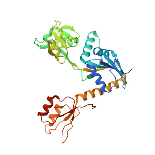
    ATP-phosphoribosyltransferase (ATP-PRT), the first enzyme of the histidine pathway, is a complex allosterically regulated enzyme, which controls the flow of intermediates through this biosynthetic pathway. The crystal structures of Escherichia coli ATP-PRT have been solved in complex with the inhibitor AMP at 2.7A and with product PR-ATP at 2.9A (the ribosyl-triphosphate could not be resolved). On the basis of binding of AMP and PR-ATP and comparison with type I PRTs, the PRPP and parts of the ATP-binding site are identified. These structures clearly identify the AMP as binding in the 5-phosphoribosyl-alpha-1-pyrophosphate (PRPP)-binding site, with the adenosine ring occupying the ATP-binding site. Comparison with the recently solved Mycobacterium tuberculosis ATP-PRT structures indicates that histidine is solely responsible for the large conformational changes observed between the hexameric forms of the enzyme. The role of oligomerisation in inhibition and the structural basis for the synergistic inhibition by histidine and AMP are discussed.


  • Organizational Affiliation

    Division of Biochemistry and Molecular Biology, Institute of Biomedical and Life Sciences, University of Glasgow, Glasgow, G12 8QQ, Scotland, UK.


Macromolecules
Find similar proteins by:  (by identity cutoff)  |  3D Structure
Entity ID: 1
MoleculeChains Sequence LengthOrganismDetailsImage
ATP phosphoribosyltransferase299Escherichia coliMutation(s): 0 
Gene Names: HISG
EC: 2.4.2.17
UniProt
Find proteins for P60757 (Escherichia coli (strain K12))
Explore P60757 
Go to UniProtKB:  P60757
Entity Groups  
Sequence Clusters30% Identity50% Identity70% Identity90% Identity95% Identity100% Identity
UniProt GroupP60757
Sequence Annotations
Expand
  • Reference Sequence
Small Molecules
Ligands 2 Unique
IDChains Name / Formula / InChI Key2D Diagram3D Interactions
PRT
Query on PRT

Download Ideal Coordinates CCD File 
B [auth A] PHOSPHORIBOSYL ATP
C15 H25 N5 O20 P4
RKNHJBVBFHDXGR-DNVSJNHSSA-N
TLA
Query on TLA

Download Ideal Coordinates CCD File 
C [auth A],
D [auth A]
L(+)-TARTARIC ACID
C4 H6 O6
FEWJPZIEWOKRBE-JCYAYHJZSA-N
Experimental Data & Validation

Experimental Data

  • Method: X-RAY DIFFRACTION
  • Resolution: 2.90 Å
  • R-Value Free: 0.272 
  • R-Value Work: 0.210 
  • R-Value Observed: 0.215 
  • Space Group: H 3 2
Unit Cell:
Length ( Å )Angle ( ˚ )
a = 132.1α = 90
b = 132.1β = 90
c = 113.379γ = 120
Software Package:
Software NamePurpose
REFMACrefinement
DENZOdata reduction
SCALEPACKdata scaling
AMoREphasing

Structure Validation

View Full Validation Report


Entry History 

Deposition Data

Revision History  (Full details and data files)

  • Version 1.0: 2004-03-02
    Type: Initial release
  • Version 1.1: 2008-04-29
    Changes: Version format compliance
  • Version 1.2: 2011-07-13
    Changes: Derived calculations, Version format compliance
  • Version 1.3: 2017-05-03
    Changes: Non-polymer description
  • Version 1.4: 2023-08-16
    Changes: Advisory, Data collection, Database references, Derived calculations, Refinement description