1PZY

W314A-BETA1,4-GALACTOSYLTRANSFERASE-I COMPLEXED WITH ALPHA-LACTALBUMIN IN THE PRESENCE OF N-ACETYLGLUCOSAMINE, UDP AND MANGANESE


Experimental Data Snapshot

  • Method: X-RAY DIFFRACTION
  • Resolution: 2.30 Å
  • R-Value Free: 0.253 
  • R-Value Work: 0.207 
  • R-Value Observed: 0.207 

wwPDB Validation   3D Report Full Report


Ligand Structure Quality Assessment 


This is version 1.6 of the entry. See complete history


Literature

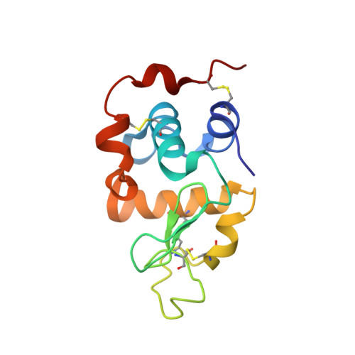
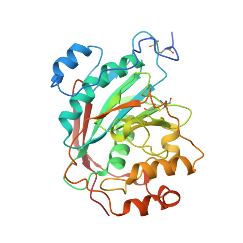
The Role of Tryptophan 314 in the Conformational Changes of beta 1,4-Galactosyltransferase-I

Ramasamy, V.Ramakrishnan, B.Boeggeman, E.Qasba, P.K.

(2003) J Mol Biol 331: 1065-1076

  • DOI: https://doi.org/10.1016/s0022-2836(03)00790-3
  • Primary Citation of Related Structures:  
    1PZT, 1PZY

  • PubMed Abstract: 

    beta1,4-Galactosyltransferase-I (beta4Gal-T1) undergoes critical conformational changes upon substrate binding from an open conformation (conf-I) to the closed conformation (conf-II). This change involves two flexible loops: the small (residues 313-316) and the long loop (residues 345-365). Upon substrate binding, Trp314 in the small flexible loop moves towards the catalytic pocket and interacts with the donor and the acceptor substrates. For a better understanding of the role played by Trp314 in the conformational changes of beta4Gal-T1, we mutated it to Ala and carried out substrate-binding, proteolytic and crystallographic studies. The W314A mutation reduces the enzymatic activity, binding to substrates and to the modifier protein, alpha-lactalbumin (LA), by over 99%. The limited proteolysis with Glu-C or Lys-C proteases shows differences in the rate of cleavage of the long loop of the wild-type and mutant W314A, indicating conformational differences in the region between the two proteins. Without substrate, the mutant crystallizes in a conformation (conf-I') (1.9A resolution crystal structure), that is not identical with, but close to an open conformation (conf-I), whereas its complex with the substrates and alpha-lactalbumin, crystallizes in a conformation (2.3A resolution crystal structure) that is identical with the closed conformation (conf-II). This study shows the crucial role Trp314 plays in the conformational state of the long loop, in the binding of substrates and in the catalytic mechanism of the enzyme.


  • Organizational Affiliation

    Structural Glycobiology Section, LECB, CCR, NCI-Frederick, Building 469, Room 221, 21702, Frederick, MD, USA.


Macromolecules
Find similar proteins by:  (by identity cutoff)  |  3D Structure
Entity ID: 1
MoleculeChains Sequence LengthOrganismDetailsImage
ALPHA-LACTALBUMIN
A, C
123Mus musculusMutation(s): 0 
UniProt
Find proteins for P29752 (Mus musculus)
Explore P29752 
Go to UniProtKB:  P29752
Entity Groups  
Sequence Clusters30% Identity50% Identity70% Identity90% Identity95% Identity100% Identity
UniProt GroupP29752
Sequence Annotations
Expand
  • Reference Sequence
Find similar proteins by:  (by identity cutoff)  |  3D Structure
Entity ID: 2
MoleculeChains Sequence LengthOrganismDetailsImage
BETA-1,4-GALACTOSYLTRANSFERASE
B, D
286Bos taurusMutation(s): 2 
EC: 2.4.1.22 (PDB Primary Data), 2.4.1.90 (PDB Primary Data), 2.4.1.38 (PDB Primary Data)
UniProt
Find proteins for P08037 (Bos taurus)
Explore P08037 
Go to UniProtKB:  P08037
Entity Groups  
Sequence Clusters30% Identity50% Identity70% Identity90% Identity95% Identity100% Identity
UniProt GroupP08037
Sequence Annotations
Expand
  • Reference Sequence
Small Molecules
Ligands 4 Unique
IDChains Name / Formula / InChI Key2D Diagram3D Interactions
UDP
Query on UDP

Download Ideal Coordinates CCD File 
H [auth B],
L [auth D]
URIDINE-5'-DIPHOSPHATE
C9 H14 N2 O12 P2
XCCTYIAWTASOJW-XVFCMESISA-N
NAG
Query on NAG

Download Ideal Coordinates CCD File 
F [auth B],
J [auth D]
2-acetamido-2-deoxy-beta-D-glucopyranose
C8 H15 N O6
OVRNDRQMDRJTHS-FMDGEEDCSA-N
MN
Query on MN

Download Ideal Coordinates CCD File 
G [auth B],
K [auth D]
MANGANESE (II) ION
Mn
WAEMQWOKJMHJLA-UHFFFAOYSA-N
CA
Query on CA

Download Ideal Coordinates CCD File 
E [auth A],
I [auth C]
CALCIUM ION
Ca
BHPQYMZQTOCNFJ-UHFFFAOYSA-N
Experimental Data & Validation

Experimental Data

  • Method: X-RAY DIFFRACTION
  • Resolution: 2.30 Å
  • R-Value Free: 0.253 
  • R-Value Work: 0.207 
  • R-Value Observed: 0.207 
  • Space Group: P 1 21 1
Unit Cell:
Length ( Å )Angle ( ˚ )
a = 55.292α = 90
b = 98.747β = 103.92
c = 102.788γ = 90
Software Package:
Software NamePurpose
DENZOdata reduction
SCALEPACKdata scaling
AMoREphasing
CNSrefinement

Structure Validation

View Full Validation Report


Ligand Structure Quality Assessment 


Entry History 

Deposition Data

Revision History  (Full details and data files)

  • Version 1.0: 2003-09-02
    Type: Initial release
  • Version 1.1: 2008-04-29
    Changes: Version format compliance
  • Version 1.2: 2011-07-13
    Changes: Non-polymer description, Version format compliance
  • Version 1.3: 2011-10-19
    Changes: Database references, Structure summary
  • Version 1.4: 2020-07-29
    Type: Remediation
    Reason: Carbohydrate remediation
    Changes: Data collection, Database references, Derived calculations, Structure summary
  • Version 1.5: 2021-10-27
    Changes: Database references, Structure summary
  • Version 1.6: 2023-08-16
    Changes: Data collection, Refinement description