1PTU | pdb_00001ptu

CRYSTAL STRUCTURE OF PROTEIN TYROSINE PHOSPHATASE 1B COMPLEXED WITH PHOSPHOTYROSINE-CONTAINING HEXA-PEPTIDE (DADEPYL-NH2)


Experimental Data Snapshot

  • Method: X-RAY DIFFRACTION
  • Resolution: 2.60 Å
  • R-Value Work: 
    0.199 (Depositor) 
  • R-Value Observed: 
    0.199 (Depositor) 

wwPDB Validation 3D Report Full Report

Validation slider image for 1PTU

This is version 1.4 of the entry. See complete history

Literature

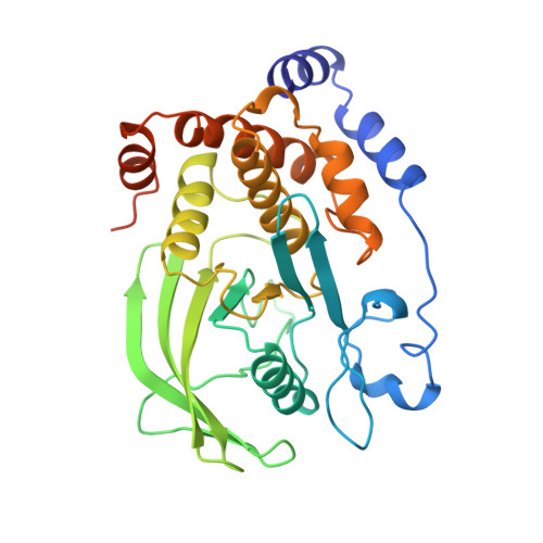
Structural basis for phosphotyrosine peptide recognition by protein tyrosine phosphatase 1B.

Jia, Z.Barford, D.Flint, A.J.Tonks, N.K.

(1995) Science 268: 1754-1758

  • DOI: https://doi.org/10.1126/science.7540771
  • Primary Citation Related Structures: 
    1PTT, 1PTU, 1PTV

  • PubMed Abstract: 

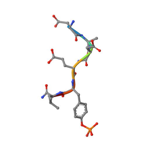
    The crystal structures of a cysteine-215-->serine mutant of protein tyrosine phosphatase 1B complexed with high-affinity peptide substrates corresponding to an autophosphorylation site of the epidermal growth factor receptor were determined. Peptide binding to the protein phosphatase was accompanied by a conformational change of a surface loop that created a phosphotyrosine recognition pocket and induced a catalytically competent form of the enzyme. The phosphotyrosine side chain is buried within the period and anchors the peptide substrate to its binding site. Hydrogen bonds between peptide main-chain atoms and the protein contribute to binding affinity, and specific interactions of acidic residues of the peptide with basic residues on the surface of the enzyme confer sequence specificity.


  • Organizational Affiliation
    • Laboratory of Molecular Biophysics, University of Oxford, UK.

Macromolecule Content 

  • Total Structure Weight: 38.17 kDa 
  • Atom Count: 2,553 
  • Modeled Residue Count: 304 
  • Deposited Residue Count: 328 
  • Unique protein chains: 2

Macromolecules

Find similar proteins by:|  3D Structure
Entity ID: 1
MoleculeChains  Sequence LengthOrganismDetailsImage
PROTEIN TYROSINE PHOSPHATASE 1B321Homo sapiensMutation(s): 1 
EC: 3.1.3.48
UniProt & NIH Common Fund Data Resources
Find proteins for P18031 (Homo sapiens)
Explore P18031 
Go to UniProtKB:  P18031
PHAROS:  P18031
GTEx:  ENSG00000196396 
Entity Groups
Sequence Clusters30% Identity50% Identity70% Identity90% Identity95% Identity100% Identity
UniProt GroupP18031
Sequence Annotations
Expand
Reference Sequence
Find similar proteins by:  Sequence   |   3D Structure  
Entity ID: 2
MoleculeChains  Sequence LengthOrganismDetailsImage
PHOSPHOTYROSINE-CONTAINING HEXA-PEPTIDE7N/AMutation(s): 0 

Small Molecules

Modified Residues  1 Unique
IDChains TypeFormula2D DiagramParent
PTR
Query on PTR
B
L-PEPTIDE LINKINGC9 H12 N O6 PTYR

Experimental Data & Validation

Experimental Data

  • Method: X-RAY DIFFRACTION
  • Resolution: 2.60 Å
  • R-Value Work:  0.199 (Depositor) 
  • R-Value Observed: 0.199 (Depositor) 
Space Group: P 31 2 1
Unit Cell:
Length ( Å )Angle ( ˚ )
a = 88.4α = 90
b = 88.4β = 90
c = 123.6γ = 120
Software Package:
Software NamePurpose
X-PLORmodel building
X-PLORrefinement
DENZOdata reduction
X-PLORphasing

Structure Validation

View Full Validation Report


Entry History 

Deposition Data

Revision History  (Full details and data files)

  • Version 1.0: 1996-08-01
    Type: Initial release
  • Version 1.1: 2008-03-24
    Changes: Version format compliance
  • Version 1.2: 2011-07-13
    Changes: Version format compliance
  • Version 1.3: 2021-11-03
    Changes: Database references, Derived calculations, Other
  • Version 1.4: 2024-10-30
    Changes: Data collection, Structure summary