1PIQ

CRYSTAL STRUCTURE OF GCN4-PIQ, A TRIMERIC COILED COIL WITH BURIED POLAR RESIDUES


Experimental Data Snapshot

  • Method: X-RAY DIFFRACTION
  • Resolution: 1.80 Å
  • R-Value Free: 0.236 
  • R-Value Work: 0.211 
  • R-Value Observed: 0.211 

wwPDB Validation   3D Report Full Report


This is version 1.3 of the entry. See complete history


Literature

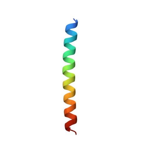
Crystal structure of GCN4-pIQI, a trimeric coiled coil with buried polar residues.

Eckert, D.M.Malashkevich, V.N.Kim, P.S.

(1998) J Mol Biol 284: 859-865

  • DOI: https://doi.org/10.1006/jmbi.1998.2214
  • Primary Citation of Related Structures:  
    1PIQ

  • PubMed Abstract: 

    Coiled coils consist of two or more alpha-helices wrapped around each other with a superhelical twist. The interfaces between helices of a coiled coil are formed by hydrophobic amino acid residues packed in a "knobs-into-holes" arrangement. Most naturally occurring coiled coils, however, also contain buried polar residues, as do the cores of the majority of naturally occurring globular proteins. Two common buried polar residues in both dimeric and trimeric coiled coils are asparagine and glutamine. In dimeric coiled coils, buried asparagine, but not glutamine, residues have been shown to confer specificity of oligomerization. We have placed a glutamine residue in the otherwise hydrophobic interior of a stable trimeric coiled coil, GCN4-pII, to study the effect of this buried polar residue in a trimeric coiled-coil environment. The resulting peptide, GCN4-pIQI, is a discrete, trimeric coiled coil with a lower stability than GCN4-pII. The crystal structure determined to 1.8 A shows that GCN4-pIQI is a trimeric coiled coil with a chloride ion coordinated by one buried glutamine residue from each monomer.


  • Organizational Affiliation

    Massachusetts Institute of Technology, Cambridge, MA, 02142, USA.


Macromolecules
Find similar proteins by:  (by identity cutoff)  |  3D Structure
Entity ID: 1
MoleculeChains Sequence LengthOrganismDetailsImage
PROTEIN (GENERAL CONTROL PROTEIN GCN4-PIQ)32N/AMutation(s): 8 
UniProt
Find proteins for P03069 (Saccharomyces cerevisiae (strain ATCC 204508 / S288c))
Explore P03069 
Go to UniProtKB:  P03069
Entity Groups  
Sequence Clusters30% Identity50% Identity70% Identity90% Identity95% Identity100% Identity
UniProt GroupP03069
Sequence Annotations
Expand
  • Reference Sequence
Small Molecules
Ligands 1 Unique
IDChains Name / Formula / InChI Key2D Diagram3D Interactions
CL
Query on CL

Download Ideal Coordinates CCD File 
B [auth A]CHLORIDE ION
Cl
VEXZGXHMUGYJMC-UHFFFAOYSA-M
Experimental Data & Validation

Experimental Data

  • Method: X-RAY DIFFRACTION
  • Resolution: 1.80 Å
  • R-Value Free: 0.236 
  • R-Value Work: 0.211 
  • R-Value Observed: 0.211 
  • Space Group: P 3 2 1
Unit Cell:
Length ( Å )Angle ( ˚ )
a = 39.66α = 90
b = 39.66β = 90
c = 47.47γ = 120
Software Package:
Software NamePurpose
AMoREphasing
CNSrefinement
DENZOdata reduction
SCALEPACKdata scaling

Structure Validation

View Full Validation Report


Entry History 

Deposition Data

Revision History  (Full details and data files)

  • Version 1.0: 1998-09-30
    Type: Initial release
  • Version 1.1: 2007-10-16
    Changes: Version format compliance
  • Version 1.2: 2011-07-13
    Changes: Derived calculations, Version format compliance
  • Version 1.3: 2021-11-03
    Changes: Database references, Derived calculations